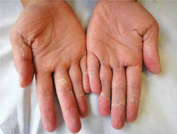
Exantemas escarlatiniformes en la infancia
DOI:
https://doi.org/10.29176/2590843X.253Resumen
Los exantemas son erupciones cutáneas de aparición súbita, generalmente asociadas a enfermedades sistémicas. Algunos de ellos comparten características clínicas con la fiebre escarlatina. Consideramos importante identificar las diferencias, principalmente de aquellas que amenazan la vida.
La presente revisión busca facilitar el diagnóstico diferencial de la fiebre escarlatina y exantemas similares como el síndrome de choque tóxico, el síndrome de piel escaldada y la enfermedad de Kawasaki, con el fin de dar un tratamiento adecuado y oportuno a cada uno según el caso.
Biografía del autor/a
Natalia Mendoza
Médica, residente de tercer año de Dermatología, Universidad Pontificia Bolivariana, Medellín, Colombia
Juliana Lopera
Médica, residente de primer año de Pediatría, Universidad Pontificia Bolivariana, Medellín, Colombia.
Ana Milena Toro
Médica dermatóloga; docente, Universidad Pontificia Bolivariana, Medellín, Colombia.
Referencias bibliográficas
2. Albisu Y. Atlas de dermatología pediátrica. Segunda edición. Enfermedades exantemáticas de la infancia. Madrid: Ergon; 2009. p. 201-31.
3. Berk DR, Bayliss SJ. MRSA, staphylococcal scalded skin syndrome, and other cutaneous bacterial emergencies. Pediatr Ann. 2010;39:627-33.
4. Harper J, Orange A, Prose N. Textbook of pediatric dermatology. Second edition. Massachusetts: Blackwell Publishing; 2006. p. 394-418.
5. Lamden KH. An outbreak of scarlet fever in a primary school. Arch Dis Child. 2011;96:394-7.
6. Sanz JC, Bascones M de LA, Martín F, Sáez-Nieto JA. Recurrent scarlet fever due to recent reinfection caused by strains unrelated to Streptococcus pyogenes. Enferm Infecc Microbiol Clin. 2005;23:388-9.
7. Hahn RG, Knox LM, Forman TA. Evaluation of poststreptococcal illness. Am Fam Physician. 2005;71:1949-54.
8. Mahajan VK, Sharma NL. Scarlet fever. Indian Pediatr. 2005;42:829-30.
9. Gidaris D, Zafeiriou D, Mavridis P, Gombakis N. Scarlet fever and hepatitis: A case report. Hippokratia. 2008;12:186-7.
10. Gómez-Carrasco JA, Lassaletta A, Ruano D. Acute hepatitis may form part of scarlet fever. An Pediatr (Barc). 2004;60:382-3.
11. Patel GK, Finlay AY. Staphylococcal scalded skin syndrome: Diagnosis and management. Am J Clin Dermatol. 2003;4:165-75.
12. Marina SS, Bocheva GS, Kazanjieva JS. Severe bacterial infections of the skin: Uncommon presentations. Clin Dermatol. 2005;23:621-9.
13. Brewer JD, Hundley MD, Meves A, Hargreaves J, McEvoy MT, Pittelkow MR. Staphylococcal scalded skin syndrome and toxic shock syndrome after tooth extraction. J Am Acad Dermatol. 2008;59:342-6.
14. Blyth M, Estela C, Young AER. Severe staphylococcal scalded skin syndrome in children. Burns. 2008;34:98-103.
15. Kikuchi K, Takahashi N, Piao C, Totsuka K, Nishida H, Uchiyama T. Molecular epidemiology of methicillin-resistant Staphylococcus aureus strains causing neonatal toxic shock syndrome-like exanthematous disease in neonatal and perinatal wards. J Clin Microbiol. 2003;41:3001-6.
16. Amagai M. Desmoglein as a target in autoimmunity and infection. J Am Acad Dermatol. 2003;48:244-52.
17. Neylon O, O’Connell NH, Slevin B, Powell J, Monahan R, Boyle L, et al. Neonatal staphylococcal scalded skin syndrome: Clinical and outbreak containment review. Eur J Pediatr. 2010;169:1503-9.
18. Kress DW. Pediatric dermatology emergencies. Curr Opin Pediatr. 2011;23:403-6.
19. Hubiche T, Bes M, Roudiere L, Langlaude F, Etienne J, Del Giudice P. Mild staphylococcal scalded skin syndrome: An underdiagnosed clinical disorder. Br J Dermatol. 2012;166:213-5.
20. The Working Group on Severe Streptococcal Infections. Defining the group A streptococcal toxic shock syndrome. Rationale and consensus definition. JAMA. 1993;269:390-1.
21. Takahashi N. Neonatal toxic shock syndrome-like exanthematous disease (NTED). Pediatr Int. 2003;45:233-7.
22. Takahashi N, Nishida H, Kato H, Imanishi K, Sakata Y, Uchiyama T. Exanthematous disease induced by toxic shock syndrome toxin 1 in the early neonatal period. Lancet. 1998;351:1614-9.
23. Chuang YY, Huang YC, Lin TY. Toxic shock syndrome in children: Epidemiology, pathogenesis, and management. Paediatr Drugs. 2005;7:11-25.
24. Hajjeh RA, Reingold A, Weil A, Shutt K, Schuchat A, Perkins BA. Toxic shock syndrome in the United States: Surveillance update, 1979-1996. Emerging Infect Dis. 1999;5:807-10.
25. Darenberg J, Ihendyane N, Sjölin J, Aufwerber E, Haidl S, Follin P, et al. Intravenous immunoglobulin G therapy in streptococcal toxic shock syndrome: A European randomized, double-blind, placebo-controlled trial. Clin Infect Dis. 2003;37:333-40.
26. Lappin E, Ferguson AJ. Gram-positive toxic shock syndromes. Lancet Infect Dis. 2009;9:281-90.
27. Shah SS, Hall M, Srivastava R, Subramony A, Levin JE. Intravenous immunoglobulin in children with streptococcal toxic shock syndrome. Clin Infect Dis. 2009;49:1369-76.
28. Rowley AH. Kawasaki disease: Novel insights into etiology and genetic susceptibility. Annu Rev Med. 2011;62:69-77.
29. Kawasaki T. Acute febrile mucocutaneous syndrome with lymphoid involvement with specific desquamation of the fingers and toes in children. Arerugi. 1967;16:178-222.
30. Puri V, Kanitkar M. Atypical Kawasaki disease. Indian J Pediatr. 2012 Fecha de consulta: 18 de junio de 2012. Mar;80(3):267-8.
31. Taubert KA, Rowley AH, Shulman ST. Nationwide survey of Kawasaki disease and acute rheumatic fever. J Pediatr. 1991;119:279-82.
32. Khubchandani RP, Viswanathan V. Pediatric vasculitides: A generalist approach. Indian J Pediatr. 2010;77:1165-71.
33. Yeung RSM. Kawasaki disease: Update on pathogenesis. Curr Opin Rheumatol. 2010;22:551-60.
34. Rowley AH, Shulman ST. Pathogenesis and management of Kawasaki disease. Expert Rev Anti Infect Ther. 2010;8:197-203.
35. Onouchi Y, Gunji T, Burns JC, Shimizu C, Newburger JW, Yashiro M, et al. ITPKC functional polymorphism associated with Kawasaki disease susceptibility and formation of coronary artery aneurysms. Nat Genet. 2008;40:35-42.
36. Macian F. NFAT proteins: Key regulators of T-cell development and function. Nat Rev Immunol. 2005;5:472-84.
37. Burgner D, Davila S, Breunis WB, Ng SB, Li Y, Bonnard C, et al. A genome-wide association study identifies novel and functionally related susceptibility loci for Kawasaki disease. PLoS Genet. 2009;5:e1000319.
38. Rowley AH, Shulman ST. Recent advances in the understanding and management of Kawasaki disease. Curr Infect Dis Rep. 2010;12:96-102.
39. Rowley AH. Incomplete (atypical) Kawasaki disease. Pediatr Infect Dis J. 2002;21:563-5.
40. Gómez-Moyano E, Vera A, Camacho J, Sanz A, Crespo-Erchiga V. Kawasaki disease complicated by cutaneous vasculitis and peripheral gangrene. J Am Acad Dermatol. 2011;64:e74-5.
41. Ducos MH, Taïeb A, Sarlangue J, Perel Y, Pedespan JM, Hehunstre JP, et al. Cutaneous manifestations of Kawasaki disease. Apropos of 30 cases. Ann Dermatol Venereol. 1993;120:589-97.
42. Mavrogeni S, Papadopoulos G, Karanasios E, Cokkinos DV. How to image Kawasaki disease: A validation of different imaging techniques. Int J Cardiol. 2008;124:27-31.
43. Newburger JW, Takahashi M, Gerber MA, Gewitz MH, Tani LY, Burns JC, et al. Diagnosis, treatment, and long-term management of Kawasaki disease: A statement for health professionals from the Committee on Rheumatic Fever, Endocarditis, and Kawasaki Disease, Council on Cardiovascular Disease in the Young, American Heart Association. Pediatrics. 2004;114:1708-33.
44. Crystal MA, Syan SK, Yeung RSM, Dipchand AI, McCrindle BW. Echocardiographic and electrocardiographic trends in children with acute Kawasaki disease. Can J Cardiol. 2008;24:776-80.
45. Lee K-Y, Rhim J-W, Kang J-H. Kawasaki disease: Laboratory findings and an immunopathogenesis on the premise of a “protein homeostasis system”. Yonsei Med J. 2012;53:262-75.
46. Newburger JW, Takahashi M, Beiser AS, Burns JC, Bastian J, Chung KJ, et al. A single intravenous infusion of gamma globulin as compared with four infusions in the treatment of acute Kawasaki syndrome. N Engl J Med. 1991;324:1633-9.
47. Newburger JW, Takahashi M, Burns JC, Beiser AS, Chung KJ, Duffy CE, et al. The treatment of Kawasaki syndrome with intravenous gamma globulin. N Engl J Med. 1986;315:341-7. Freeman AF, Shulman ST. Refractory Kawasaki disease. Pediatr Infect Dis J. 2004;23:463-4.
48. Furukawa T, Kishiro M, Akimoto K, Nagata S, Shimizu T, Yamashiro Y. Effects of steroid pulse therapy on immunoglobulinresistant Kawasaki disease. Arch Dis Child. 2008;93:142-6.
49. Burns JC, Best BM, Mejias A, Mahony L, Fixler DE, Jafri HS, et al. Infliximab treatment of intravenous immunoglobulin-resistant Kawasaki disease. J Pediatr. 2008;153:833-8.
Cómo citar
Descargas

Descargas
Publicado
Cómo citar
Número
Sección

| Estadísticas de artículo | |
|---|---|
| Vistas de resúmenes | |
| Vistas de PDF | |
| Descargas de PDF | |
| Vistas de HTML | |
| Otras vistas | |


