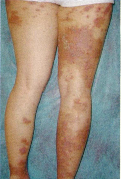
Dermatosis eosinofílicas
Palabras clave:
Eosinófilo, sindrome de Wells, sindrome de Shulman, granuloma facial, síndrome de hipereosinofiliaResumen
La presencia de eosinófilos es un hallazgo útil que conduce al diagnostico de varias enfermedades cutáneas. En ocaciones ellos son indespecíficos y requieresn de una estricta correlación clínica.
En este artículo se revisa información acerca de la patogénesis, presentación clínica, diagnóstico y tratamiento de las patologías que característicamente cursan con eosinófilos a nivel tisular.
Biografía del autor/a
Sara González Trujillo, Universidad Pontificia Bolivariana
Residente III Dermatología de la Universidad Pontificia Bolibariana. Medellin - Colombia.
Luz Marina Gómez Vargas, Universidad Pontificia Bolivariana
Dermatóloga - Asesora de la Universidad Pontificia Bolivariana. Medellín - Colombia.
Rodrigo Restrepo Molina, Universidad Pontificia Bolivariana
Patólogo - Asesor de la Universidad Pontificia Bolivariana. Medellín - Colombia.
Referencias bibliográficas
2. Weedon D. Skin Pathology. Ed Churchill Livingstone London;2002: 1059-1061.
3. Rothenberg M. Eosinophilia. New England Journal of Medicine 1998; 338:1592-1600.
https://doi.org/10.1056/NEJM199805283382206
4. Fench L, Shapiro M, Junkins-Hopkins M, Wolfe J, Rook A. Eosinphilic fasciitis and eosinophilic cellulites in a patient with abnormal circulating clonal T cells: increased production of interleukin 5 and inhibition by interferon alfa. Am J Dermatol 2003; 49:1170-1174.
https://doi.org/10.1016/S0190-9622(03)00447-X
5. España A, Sanz E, Sola B, Gil H. Wells síndrome (eosinophilc cellulitis) : correlation between clinical activity, eosinophil levels, eosinophil cation protein and interleukin 5.Br J Dermatol 1999;140:127-130.
https://doi.org/10.1046/j.1365-2133.1999.02621.x
6. Moossavi M, Mehergan D. Wells syndrome: a clinical and histopathologic review of seven cases. lnt J Der matol 2003;42:62-67.
https://doi.org/10.1046/j.1365-4362.2003.01705.x
7. Stetson C. Dermatosis eosinofílicas. En Bolonia J, Jorizzo J, Rapini R, Horn T, Mascaró J, Mancicni A, Sa lashe S, Saurat J, Swing G eds. Dermatología, primera edición. Madrid, España: Elsevier;2004:403-409.
8. Ludwing R, Grundmann-Kollman M, Holtmeier W, Glas J, Podota M, Kaufmann R, Zollner T. Herpes simple virus type 2 associated eosinophilic cellulitis. Am J Dermatol 2003; 48S60-1.9.
https://doi.org/10.1067/mjd.2003.20
9. GhilslainP,EeckhoutV.Eosinophiliccellulitisofpapu lonodular presentation. JEADV 2005; 19:226-227.
https://doi.org/10.1111/j.1468-3083.2005.01027.x
10. SchuttelaarM,JonkmanM.Bullouseosinophiliccellu lites associated with Churg Strauss syndrome. JEADV 2003;17:91-93.
https://doi.org/10.1046/j.1468-3083.2003.00648.x
11. Sommer S, Wilkinson S, Merchant W. Eosinophilic cellulites following the lines of Blaschko. Clin Exp Dermatol 1999; 24:449-451.
https://doi.org/10.1046/j.1365-2230.1999.00529.x
12. Mckee P, Calonge E, Granter S. Pthology of the skin with Clinical Correlation. Ed Elsevier, China;2005;696- 702.
13. Hish K, Ludwing R, Wolter M, Zolliner T, lhardt K, Kaufmann R, Boehnicke W. Eosinophilic cellulites as sociated with colon carcinoma. Journalder Deutschen Dermatologis Chen Cesellschaft 2005; 3:530-1.
https://doi.org/10.1111/j.1610-0387.2005.05726.x
14. Fujii K, Tanabe H, Kanno Y, Konishi K, Ohgou N. Eosinophilic cellulitis as a cutaneus manifestation of idiopatic hypereosinophilic síndrome. Am J Dermatol 2003; 49:1174-7.
https://doi.org/10.1016/S0190-9622(03)00466-3
15. ChanV,SoansB,MathersD.Ultrasoundandmagnetic resonance imaging features in a patient with eosnophilic fasciitis. Australasian Radiology 2004; 48:414-417.
https://doi.org/10.1111/j.0004-8461.2004.01331.x
16. Schiener R, Behrens- Williams S, Gottloeber P, Pille- kamp H, Peter R, Kerscher M. Eosinophilic fasciitis treated with psoralen-ultraviolet a bath photochemo- therapy. Br J Dermatol 200; 142:804-807.
https://doi.org/10.1046/j.1365-2133.2000.03431.x
17. Romano C, Rubegni P, De Aloe G, Stanghellini E, Do Ascenzo L, Andreassi L, Fimiani M. Extracorporeal photochemotherapy in the treatment of eosinophilic fasciitis. JEADV 2003; 17:10-13.
https://doi.org/10.1046/j.1468-3083.2003.00587.x
18. Valencia C, Chang A, Kirsner R, Kerdel F. Eosinophilic fasciitis responsive to treatment with pulsed esteroids and cyclosporine. lnter J Dermatol 1999; 38 (5) 367.
https://doi.org/10.1046/j.1365-4362.1999.00695.x
19. Jinnin M, lhn H, Yamane K, Asano Y, Yazawa N, Tamaki K. Clinical and laboratory investigations serum levels of tissue inhibitor of metalloproteinase-1 and 2 in pa- tients with eosinophilic fasciitis. Br J Dermatol 2004; 151:407-412.
https://doi.org/10.1111/j.1365-2133.2004.06062.x
20. Zargari O. Disseminated granuloma faciale. lnt J Der- matol 2004; 43:208-21O.
https://doi.org/10.1111/j.1365-4632.2004.02099.x
21. Dowlati B, Alireza B, DowlatiY. Granuloma faciale: successful treatment of nine cases with a combination of cryoterapy and intralesional corticosteroid inyection. lnt J Dermatol 1997; 36:548-551.
https://doi.org/10.1046/j.1365-4362.1997.00161.x
22. Roussaki-Schulze A, Klimi E, Zaafiriou E, Aroni K, Mpaltopoulos A, Aravantinos E, Mellekos M. Granu- loma faciale associated with adenocarcinoma of the prostate. lnt J Dermatol 2002; 41:901-903.
https://doi.org/10.1046/j.1365-4362.2002.01646_1.x
23. lnanir 1, Alvur Y. Granuloma faciale with extrafacial lesions. Br J Dermatol 2001; 145:360.
https://doi.org/10.1046/j.1365-2133.2001.04360.x
24. Yung A, Wachsmuth R, Ramnath R, Merchant W, Myatt A, Sheehan-Dare R. Eosinophilic angiocentric fibrosis- a rare variant of granuloma faciale which may present to the dermatologist. Br J Dermatol 2005; 152:565-567.
https://doi.org/10.1111/j.1365-2133.2005.06439.x
25. Selvaag E, Roalda B. lmmunohistochemical findings in granuloma faciale. The role of eosinophilic granulo- cytes. JEADV 2000; 14:513-522.
https://doi.org/10.1046/j.1468-3083.2000.00156-4.x
26. Ludwing E, Allam J, Bieber T, Novak N. New treatment modalities for granuloma faciale. Br J Dermatol 2003; 149:634-637.
https://doi.org/10.1046/j.1365-2133.2003.05550.x
27. Holme S, Laidler P, Holt P Concurrent granuloma faciale and eosinophilic angiocentric fibrosis. Br J Dermatol 2005; 153:842-868.
https://doi.org/10.1111/j.1365-2133.2005.06864.x
28. Brito-BabapulleF.Review:Theeosinophilias,incluging the idiopatic hypereosinophilic syndrome. Br J Haema- tol 2003; 121:203-223.
https://doi.org/10.1046/j.1365-2141.2003.04195.x
29. Cogan E, Schandene L, Crusiaux A, Cochaux P, Velu T, Goldman M. Clona! proliferation of type 2 helper T cells in a man with the hypereosinophilic syndrome. N Engl J Med 1994; 330:535-538.
https://doi.org/10.1056/NEJM199402243300804
30. Tsunemi Y, ldezuki T, Nakamura K, Tamaki K. Termal endotelial cells express eotaxin in hypereosinphilic síndrome. J Acad Dermatol 2003; 49:918-921.
https://doi.org/10.1016/S0190-9622(03)00449-3
31. RoufosseF,CoganE,GoldmanM.Thehypereosophilic síndrome. Ann Rev Med 2003; 54:169-184.
https://doi.org/10.1146/annurev.med.54.101601.152431
32. Gleich G, Leiferman K, Tefferi J, Pardanani A, But- terfield J. Treatment of hypereosinophilic syndrome with imatinib mesilate. Lancet 2002; 359:1577- 1578.
https://doi.org/10.1016/S0140-6736(02)08505-7
33. WerchniakA,PerryA,DinulosJ.Eosinophilic,polymor- phic, and pruritic eruption associated with radiotherapy (EPPER) in a patient with breast cancer. J Am Acad Dermatol 2006;54:728-729.
https://doi.org/10.1016/j.jaad.2005.02.038
34. Rueda R, Valencia 1, Covelli C, Escobar C, Alzate A, Saldarriaga B, Sanclemente G, Blank A, Falabella R. Eosinophilic, polymorphic, and pruritic eruption as- sociated with radiotherapy. Arch Dermatol 1999;135 (7):804-81o.
https://doi.org/10.1001/archderm.135.7.804
Cómo citar
Descargas

Descargas
Publicado
Cómo citar
Número
Sección
| Estadísticas de artículo | |
|---|---|
| Vistas de resúmenes | |
| Vistas de PDF | |
| Descargas de PDF | |
| Vistas de HTML | |
| Otras vistas | |


