
Lesión pigmentada de Talón
Resumen
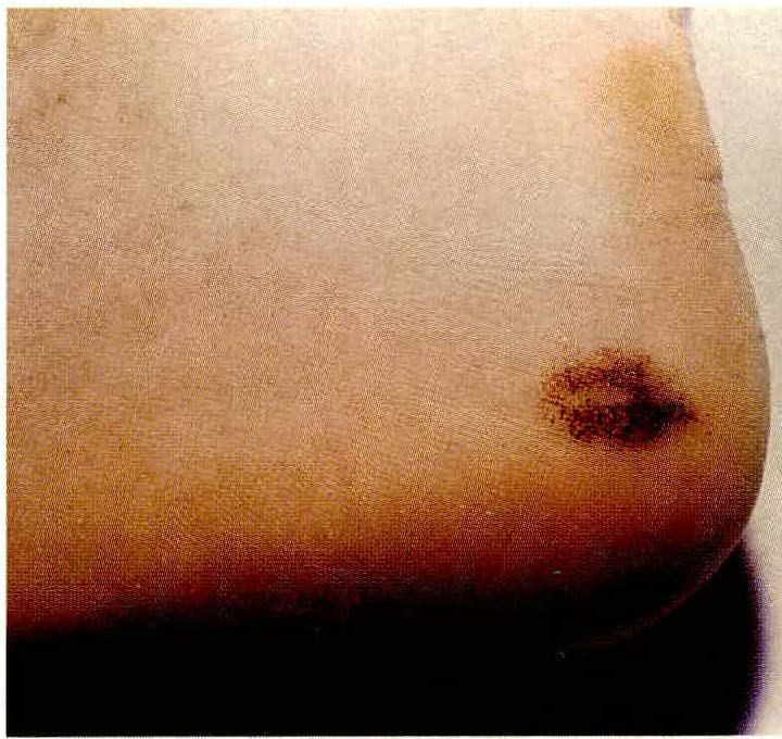
Se reporta el caso de un paciente que presentaba una mácula café oscura en el talón. Se le realiza biopsia y se discute su diagnóstico diferencial.
Biografía del autor/a
Gina Evelyn Gómez aya, Hospital Militar Central
Servicio de Dermatología, Hospital Militar Central, Santafé de Bogotá.
Gerzain Rodríguez, Universidad Nacional
Profesor Titular, Laboratorio de Patología, Instituto Nacional de Salud, A.A. 80334, Facultad de Medicina, Universidad Nacional, Santafé de Bogotá, Colombia.
Juan Guillermo Chalela, Escuela Colombiana de Medicina
Servicio de Dermatología, Escuela Colombiana de Medicina, Santafé de Bogotá.
Referencias bibliográficas
1. Fitzpatrick T., Eisen A., WolffK. et al. Dover JS., Talon Noir. Sports Dermatology .. Dermatology in General Medicine. 4th ed. Me Graw Hill, Lnc. 1993; pp: 1617-1618.
2. Rook, Wilkinson, Ebling. Black Heel. Text Book o/ Dermatology. 5th electronic de. Champion, Burton & Ebling (eds). 1992.
3. Ayres S, Mihan R. Calcaneal petechiae. Arch Dermatol. 1972; 106-262.
https://doi.org/10.1001/archderm.1972.01620110086022
4. Yafee H. Talon noir. Arch Dermatol. 1971; 104: 452.
https://doi.org/10.1001/archderm.104.4.452b
5. lzumi S, Mihan R. Calcaneal petechiae. Arch Dermalol. 1974; 109-261.
https://doi.org/10.1001/archderm.1974.01630020069019
6. Saida T, Oguchi S, Oshihara Y. In vivo observat_ion of magnified features ofpigmented lesions on volar skm using video macroscope. Arch Dermatol. 1995: 131: 298-304.
https://doi.org/10.1001/archderm.131.3.298
7. Hafner, J., Haenseler, E., Ossent, P, et al. Benzidine stain for histochemical detection ofhemoglobin in splinter hemorrage and black heel. Am J Dermatopath. 1995; 17: 362- 367.
https://doi.org/10.1097/00000372-199508000-00010
8. Mehregan A., Mehregan D., Hashimoto K. et al. Black Heel. Pinkus' Guide for Dermatohistopathology. En: Pigmentary Disorders. 61h ed. Appleton & Lange, orwalk 1995, pp 447.
9. Okun M., Eldestein L, Fisher B. Gross and microscopic pathology of the skin. Black heel. En: Sorne non melanotyc lesions clinically resembling malignan! melanoma. 2nd ed. 1994. Demiatopathology F oundation Press, lnc. pp 1258.
Cómo citar
1.
Gómez aya GE, Rodríguez G, Chalela JG. Lesión pigmentada de Talón. rev. asoc. colomb. dermatol. cir. dematol. [Internet]. 1 de junio de 1999 [citado 3 de julio de 2024];7(2):79-80. Disponible en: https://revista.asocolderma.org.co/index.php/asocolderma/article/view/765
Descargas
Los datos de descargas todavía no están disponibles.
Descargas
Publicado
1999-06-01
Cómo citar
1.
Gómez aya GE, Rodríguez G, Chalela JG. Lesión pigmentada de Talón. rev. asoc. colomb. dermatol. cir. dematol. [Internet]. 1 de junio de 1999 [citado 3 de julio de 2024];7(2):79-80. Disponible en: https://revista.asocolderma.org.co/index.php/asocolderma/article/view/765
Número
Sección
Minicasos

| Estadísticas de artículo | |
|---|---|
| Vistas de resúmenes | |
| Vistas de PDF | |
| Descargas de PDF | |
| Vistas de HTML | |
| Otras vistas | |


