
¿Reconoce esta clave diagnóstica?
Parte II
Keywords:
Granuloma annulare, necrobiotic disordersAbstract
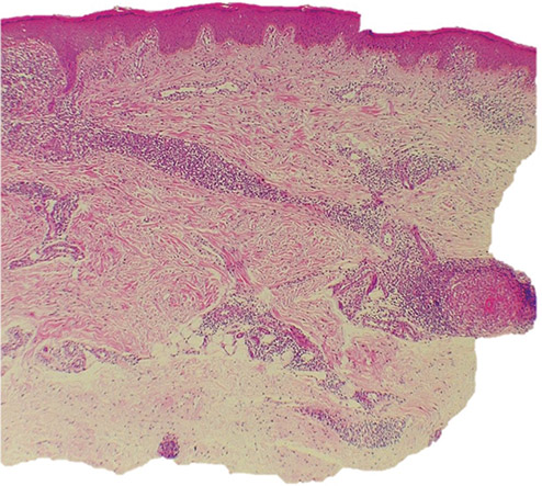
We present the case of a 38-year-old woman who presents with a 1 year evolution erythematous macula in the right heel without improvement with topical treatments. On inspection, the lesion is an erythematous plaque with an annular appearance. Histopathology reveals palisading granulomas, foci of necrobiosis and a perivascular granuloma.
The presence of confluent necrobiosis with extension to the subcutaneous cellular tissue with palisading granulomas in the absence of the sign of the sandwich, are the diagnostic key of the annular granuloma. Additionally, we observed a rare finding: a medium-sized vessel with perivascular granuloma formation, which corresponds to a granulomatous vasculitis, a morphological sign seen almost exclusively in the annular granuloma.
Author Biographies
Isabel Flórez
Médica, residente de Patología, Universidad de Antioquia, Medellín, Colombia
Saúl Rivero
Médico, residente de Patología, Universidad de Cartagena, Cartagena, Colombia
Luis Correa
Médico dermatopatólogo; profesor de Dermatopatología, Sección de Dermatología, Centro de Investigaciones Dermatológicas CIDERM, Facultad de Medicina, Universidad de Antioquia, Medellín, Colombia
References
Haim S, Shafrir A, Haim N, Lichtig C. Microangiopathy in cases of granuloma annulare. Dermatologica. 1973;147:261-6.
https://doi.org/10.1159/000251878
Haim S, Friedman-Birnbaum R, Shafrir A. Generalized granuloma annulare: Relationship to diabetes mellitus as revealed in 8 cases. Br J Derm. 1970;83:302-5.
https://doi.org/10.1111/j.1365-2133.1970.tb15704.x
Dahl MV, Ullman S, Goltz RW. Vasculitis in granuloma annulare; Histopathology and direct immunofluorescence. Arch Dermatol. 1977;113:463-7.
https://doi.org/10.1001/archderm.1977.01640040071010
Magro CM, Crowson AN, Regauer S. Granuloma annulare and necrobiosis lipoidica tissue reactions as a manifestation of systemic disease. Hum Pathol. 1996;27:50-6.
https://doi.org/10.1016/S0046-8177(96)90137-9
Günes P, Göktay F, Mansur AT, Köker F, Erfan G. Collagen-elastic tissue changes and vascular involvement in granuloma annulare: a review of 35 cases. J Cutan Pathol. 2009;36:838-44.
https://doi.org/10.1111/j.1600-0560.2008.01169.x
Ko CJ, Glusac EJ. Noninfectious granulomas. In: Elder DE, editor. Lever's Histopathology of the Skin. 11th edition. Philadelphia: Wolters Kluwer; 2015. p. 427-31.
Patterson JW. The granulomatous reaction pattern. In: Patterson JW, Hosler GA, editors. Weedon's Skin Pathology. 4th edition. Charlottesville, VA: Churchill Livingstone Elsevier; 2016. p. 189-218.
How to Cite
Downloads
Downloads
Published
How to Cite
Issue
Section

| Article metrics | |
|---|---|
| Abstract views | |
| Galley vies | |
| PDF Views | |
| HTML views | |
| Other views | |


