
¿Reconoce esta clave diagnóstica?
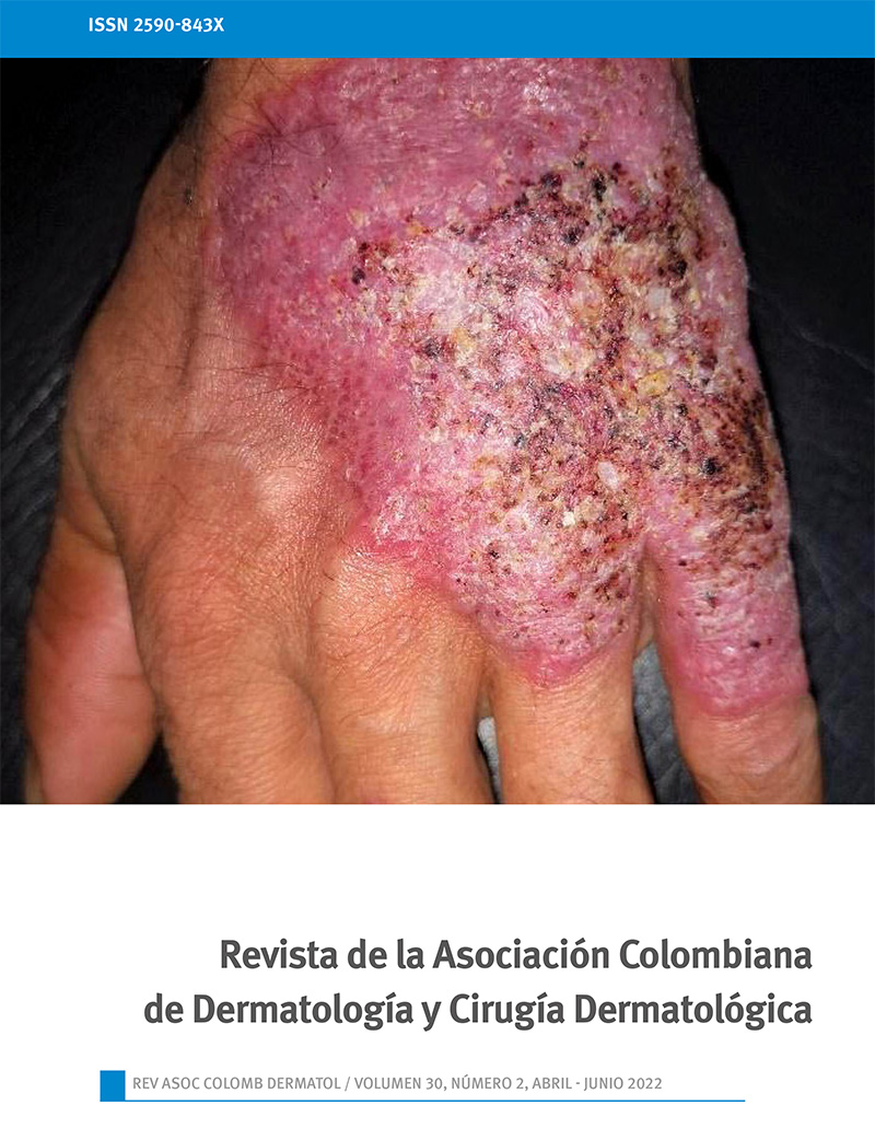
Woman with plaques and erosions on photoexposed skin
DOI:
https://doi.org/10.29176/2590843X.1607Keywords:
Cutaneous lupus erythematosus; Stevens Johnson syndrome; Systemic lupus erythematosus; Toxic epidermal necrolysisAbstract
A middle-aged woman presented with immunological, mucocutaneous, hematological and renal signs and symptoms, with a final diagnosis of systemic lupus erythematosus with toxic epidermal necrolysis (TEN) type manifestation. Systemic lupus erythematosus is accompanied by multiple dermatological manifestations, of these, different forms of bullous lesions have been described, including lupus erythematosus with TEN-type manifestation, an entity with lower morbidity and mortality than classic TEN and which normally
presents an excellent response to systemic corticosteroids. The presentation of this clinical case is considered relevant due to its rarity and because it can be an important clue for the diagnosis of systemic lupus erythematosus.
References
Chanprapaph K, Tankunakorn J, Suchonwanit P, Rutnin S. Dermatologic Manifestations, Histologic Features and Disease Progression among Cutaneous Lupus Erythematosus Subtypes: A Prospective Observational Study in Asians. Dermatol Ther (Heidelb). 2021;11(1):131-47. https://doi.org/10.1007/s13555-020-00471-y
de Risi-Pugliese T, Cohen Aubart F, Haroche J, Moguelet P, Grootenboer-Mignot S, Mathian A, et al. Clinical, histological, immunological presentations and outcomes of bullous systemic lupus erythematosus: 10 New cases and a literature review of 118 cases. Semin Arthritis Rheum. 2018;48(1):83-9. https://doi.org/10.1016/j.semarthrit.2017.11.003
Rutnin S, Chanprapaph K. Vesiculobullous diseases in relation to lupus erythematosus. Clin Cosmet Investig Dermatol. 2019;12:653-67. https://doi.org/10.2147/CCID.S220906
Auquier-Dunant A, Mockenhaupt M, Naldi L, Correia O, Schröder W, Roujeau JC, et al. Severe Cutaneous Adverse Reactions. Correlations between clinical patterns and causes of erythema multiforme majus, Stevens-Johnson syndrome, and toxic epidermal necrolysis: results of an international prospective study. Arch Dermatol. 2002;138(8):1019-24. https://doi.org/10.1001/archderm.138.8.1019
Ziemer M, Kardaun SH, Liss Y, Mockenhaupt M. Stevens-Johnson syndrome and toxic epidermal necrolysis in patients with lupus erythematosus: A descriptive study of 17 cases from a national registry and review of the literature. Br J Dermatol. 2012;166(3):575-600.
Mandelcorn R, Shear NH. Lupus-associated toxic epidermal necrolysis: a novel manifestation of lupus? J Am Acad Dermatol. 2003;48(4):525-9. https://doi.org/10.1067/mjd.2003.107
Tankunakorn J, Sawatwarakul S, Vachiramon V, Chanprapaph K. Stevens-Johnson Syndrome and Toxic Epidermal Necrolysis-Like Lupus Erythematosus. J Clin Rheumatol. 2019;25(5):224-31. https://doi.org/10.1097/RHU.0000000000000830
Ting W, Stone MS, Racila D, Scofield RH, Sontheimer RD. Toxic epidermal necrolysis-like acute cutaneous lupus erythematosus and the spectrum of the acute syndrome of apoptotic pan-epidermolysis (ASAP): a case report, concept review and proposal for new classification of lupus erythematosus vesiculobullous skin lesions. Lupus. 2004;13(12):941-50. https://doi.org/10.1191/0961203304lu2037sa
Wenzel J, Tüting T. An IFN-associated cytotoxic cellular immune response against viral, self-, or tumor antigens is a common pathogenetic feature in “interface dermatitis”. J Invest Dermatol. 2008;128(10):2392-402. https://doi.org/10.1038/jid.2008.96
Boontaveeyuwat E, Silpa-archa N, Kulthanan K. Toxic epidermal necrolysis-like acute cutaneous lupus erythematosus (TEN-like ACLE) in SLE patients: a report of two cases. Asian Pac J Allergy Immunol. 2012;30(1):83-7.
Torchia D, Romanelli P, Kerdel FA. Erythema multiforme and Stevens-Johnson syndrome/toxic epidermal necrolysis associated with lupus erythematosus. J Am Acad Dermatol. 2012;67(3):417-21. https://doi.org/10.1016/j.jaad.2011.10.012
Zargham H, Ghazal S, Watters K, Nguyen KH. A case of toxic epidermal necrosis-like cutaneous eruption as the first manifestation and clue to the diagnosis of systemic lupus erythematosus: A case report. SAGE Open Med Case Rep. 2020;8:2050313X20940420. https://doi.org/10.1177/2050313X20940420
How to Cite
Downloads
Downloads
Published
How to Cite
Issue
Section
License
Copyright (c) 2023 Revista de la Asociación Colombiana de Dermatología y Cirugía Dermatológica

This work is licensed under a Creative Commons Attribution-NonCommercial-ShareAlike 4.0 International License.

| Article metrics | |
|---|---|
| Abstract views | |
| Galley vies | |
| PDF Views | |
| HTML views | |
| Other views | |


