
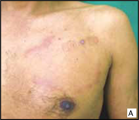
Palpable migratory arciform lesions.
Keywords:
chronic cutaneous lupus erythematosus, lupus erythematosus tumidus, Jessner-Kanof lymphocytic infiltrationAbstract
A 56-year-old man presented with an elevated arciform, annular, erythematous plaque of thirty years on the left side of his upper trunk. The lesion had shown spontaneous resolution with the formation of new arciform or semiannular lesions.
The histological findings showed the epidermis unchanged, in the dermis a dense superficial and deep lymphocytic infiltration, perivascular and periadnexal; Alcian blue staining revealed a significant amount of mucin between collagen bundles, which was negative for Borrelia spp. Immunohistochemistry stains showed than the majority of lymphocytes had a T-cell phenotype, CD3+ CD5+ positive, and CD30- negative. Similar histopathological findings are seen in other pathologies such as Jessner-Kanof lymphocytic infiltration, erythema annular centrifugum, Borrelia spp. infection, polymorphic light eruption, reticular erythematous mucinosis or pseudolymphoma, and, consequently, histological differentiation may be difficult. This case with nonscarring, erythematous, succulent plaques with no surface changes over sun exposed areas, perivascular and periadnexal superficial and deep lymphocytic infiltration, and interstitial mucin deposition is charactetistic of lupus erythematosus tumidus.
Author Biographies
Lucy García
Médica dermatóloga, M.Sc. Ciencias Básicas con énfasis en Inmunología; docente de Dermatología, Universidad del Valle, Cali, Colombia
Sara Lozada
Médica residente, tercer año de Dermatología, Universidad del Valle, Cali, Colombia
Liliana Muñoz
Médica dermatóloga, dermatopatóloga; docente de Dermatología, Universidad del Valle, Cali, Colombia.
References
2. Gougerot H, Burnier M. Lupus erythematosus tumidus. Bull Soc Fr Derm Syph. 1930;12:91.
3. Kuhn A, Sonntag M, Richter-Hintz D, Oslislo C, Megahed M, Ruzicka T, et al. Phototesting in lupus erythematosus tumidus: review of 60 patients. Photochem Photobiol. 2001;73:532-6.
4. Teixeira M, Ferreira M, Alves R, Selores M. Lupus erythematosus tumidus: an underestimated entity. Lupus. 2006;15:296-300.
5. Kuhn A, Richter-Hintz D, Oslislo C, Ruzicka T, Megahed M, Lehmann P. Lupus erythematosus tumidus–a neglected subset of cutaneous lupus erythematosus: report of 40 cases. Arch Dermatol. 2000;136:1033-41.
6. Vieira V, Del Pozo J, Yebra-Pimentel MT, Martínez W, Fonseca E. Lupus erythematosus tumidus: a series of 26 cases. Int J Dermatol. 2006;45:512-7.
7. Obermoser G, Schwingshackl P, Weber F, Stanarevic G, Zelger B , Romanin N, Sepp N. Recruitment of plasmacytoid dendritic cells in ultraviolet irradiation-induced lupus erythematosus tumidus. Br J Dermatol. 2009;160:197-200.
8. Rémy-Leroux V, Léonard F, Lambert D, Wechsler J, Cribier B, Thomas P, et al. Comparison of histopathology and clinical characteristics of Jessner’s lymphocytic infiltration of the skin and lupus erythematosus tumidus: Multicenter study of 46 cases. J Am Acad Dermatol. 2008;58:217-23.
9. Rongioletti F, Rebora A. Cutaneous mucinoses: microscopic criteria for diagnosis. Am J Dermatopathol. 2001;23:257-67.
10. Albrecht J, Fine LA, Piette W. Drug-associated lymphoma and pseudolymphoma: recognition and management. Dermatol Clin. 2007;25:233-44.
11. Grange F, Wechsler J, Guillaume JC, Tortel J, Tortel MC, Audhuy B, et al. Borrelia burgdorferi–associated lymphocytoma cutis simulating a primary cutaneous large B-cell lymphoma. J Am Acad Dermatol. 2002;47:530-4.
How to Cite
Downloads
Downloads
Published
How to Cite
Issue
Section

| Article metrics | |
|---|---|
| Abstract views | |
| Galley vies | |
| PDF Views | |
| HTML views | |
| Other views | |


