
Spectral analysis of acquired melanocytic lesions
DOI:
https://doi.org/10.29176/2590843X.309Keywords:
Nevus pigmented, spectrum analysis, biopsyAbstract
Introduction: History has established different methods for morphological information of pigmented lesions. The gold standard for diagnosis is biopsy; however, noninvasive optical techniques such as diffuse spectrophotometry reflectance have been developed.
Objective: To characterize acquired melanocytic lesions using diffuse spectrophotometry reflectance.
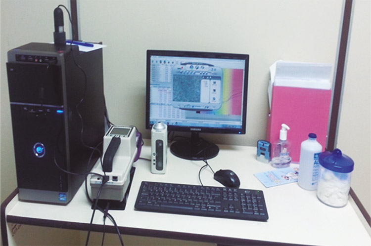
Materials and methods: We carried out a prospective study, cohort study. The population consisted of 63 patients with acquired melanocytic lesions who consulted the Service of Dermatology, Ciudad Hospitalaria Dr. Enrique Tejera, Valencia, Venezuela. The sampled included 120 nevi classified clinical and histopathological diagnosis. For data collection a Hunterlab MiniScan XE Plus spectrophotometer was used.
Results: There are differences in reflectance between each of the lesions studied. Dysplastic nevi present greater melanin absorption which is related to a deeper lesion on the skin.
Conclusion: Melanocytic nevi studied are characterized by a defined spectral signature. It is recommended to include diffuse spectrophotometry reflectance, as an optional method of diagnosis and monitoring in dermatologic consultation.
Author Biographies
Iriana Álvarez
Médica dermatóloga, Ciudad Hospitalaria Dr. Enrique Tejera, Facultad de Ciencias de la Salud, Universidad de Carabobo, Valencia, Venezuela
Sandra Vivas
Médica dermatóloga; jefe, coordinadora de posgrado de Dermatología, Universidad de Carabobo, Valencia, Venezuela
Aarón Muñoz
Licenciado en Física, maestría y doctorado en Ciencias Básicas con especialidad en Óptica; director general, Centro de Investigaciones Médicas y Biotecnológicas, Universidad de Carabobo, Valencia, Venezuela
References
Rex J, Ferrandiz C. Nevus melanocíticos. Fecha de consulta: 4 de febrero de 2015. Disponible en: https://www.aeped.es/sites/default/files/documentos/nevus.pdf
Friedman R. Rigel DS, Kopf AW. Early detection of malignant melanoma: The role of physician examination and self examination of the skin. CA Cancer J Clin. 1985;35:130-51.
https://doi.org/10.3322/canjclin.35.3.130
Wolf K, Johnson R. Color atlas & synopsis of clinical dermatology. Buenos Aires: McGraw Hill; 2009. p. 178-91.
Wolff K, Goldsmith L, Katz S, Gilchrest B, Paller A, Leffell D. Fitzpatrick Dermatología en Medicina General. Séptima edición. Buenos Aires: Editorial Panamericana; 2010. p. 1099-133.
Garnacho GM, Moreno JC. Trastornos de la pigmentación: lentigos, nevus y melanoma. Fotoprotección. Servicio de Dermatología. Hospital Universitario Reina Sofía de Córdoba. Pediatr Integral. 2012;XVI: 321-31.
Lacarrubba F. Use of videodermatoscopy in dermatology. En: Berardesca E, Maibach H, Wilheim K, editores. Non Invasive Diagnostic Techniques in Clinical Dermatology. Berlin: Springer; 2014. p. 3-26.
https://doi.org/10.1007/978-3-642-32109-2_1
Bono A, Tomatis S, Bartoli C, Tragni G, Radaelli G, Maurichi A, et al. The ABCD system of melanoma detection. A spectrophotometric analysis of the asymmetry, border, color and dimension. Cancer, 1999;85:72-7.
https://doi.org/10.1002/(SICI)1097-0142(19990101)85:1<72::AID-CNCR10>3.0.CO;2-Q
Fodor L, Ullmann Y, Elman M. Aplicaciones estéticas de la luz pulsada intensa. Israel. Amolca; 2012. p. 11- 20
Cordo M, Sendra J, Viera A, Santana A, López SM. Diferenciación de piel sana y lesiones cutáneas pigmentadas mediante espectroscopía de reflectancia óptica difusa. Óptica Pura y Aplicada. 2006;39:341-54.
Orozco E. Estudio de lesiones en piel mediante espectroscopía de reflexión difusa (tesis). Puebla, México: Instituto Nacional de Astrofísica, Óptica y Electrónica; 2009.
Tomatis S, Bono A, Bartoli C, Carrara M, Lualdi M, Tragni G, et al. Automated melanoma detection: multispectral imaging and neural network approach for classification. Phys Med. 2003;30:212-21.
https://doi.org/10.1118/1.1538230
Marchesini R, Brambilla M, Clemente C, Maniezzo M, Sichirollo AE, Testori A, et al. In vivo spectrophotometric evaluation of neoplastic and nonneoplastic skin pigmented lesions. III. CCD camera-based reflectance imaging. Photochem Photobiol. 1995;62:151-4.
https://doi.org/10.1111/j.1751-1097.1995.tb05251.x
Scarisbrick J, Pickard C, Lee A, Briggs G, Johnson K, Bown S, et al. Elastic scattering spectroscopy in the diagnosis of pigmented lesions: Comparison with clinical and histopathological diagnosis. Proceedings of SPIE 2003,5141:147-56.
https://doi.org/10.1117/12.501181
Marchesini R, Cascinelli N, Bambrilla M, Clemente C, Mascheroni L, Pignoli E, et al. In vivo spectrophotometric evaluation of neoplastic and nonneoplastic skin pigmented lesions. II. Discriminant analysis between nevus and melanoma. Photochem Photobiol. 1992;55:515-22.
https://doi.org/10.1111/j.1751-1097.1992.tb04272.x
Orozco E, Iruretagoyena G, Vázquez S, Delgado J, Castro J, Gutiérrez F. Métodos de clasificación para identificar lesiones en piel a partir de espectros de reflexión difusa. Revista Ingeniería Biomédica. 2010;4:34-40.
Ascierto P, Palla M, Ayala F, De Michele I, Caracò C, Daponte A, et al. The role of spectrophotometry in the diagnosis of melanoma. 2010;10:5.
https://doi.org/10.1186/1471-5945-10-5
Zonios G, Dimou A. Optical properties of human melanocytic nevi in vivo. Photochem Photobiol. 2009;85:298-303.
https://doi.org/10.1111/j.1751-1097.2008.00436.x
Feinsilber D, Acosta A, Rosati O, Kogan N Schroh R, Corbella C. Método de codificación de lesiones pigmentarias. Dermatol Argent. 2014;20:94-8.
García A, Smith E, Zou J, Duvic M, Prieto V, Wang LV. In-vivo characterization of optical properties of pigmented skin lesions including melanoma using oblique incidence diffuse reflectance spectrometry. J Biomed Opt. 2011;16:020501.
https://doi.org/10.1117/1.3536509
Carnero-Pardo C. Evaluación de las pruebas diagnósticas. Rev Neurol. 2005;40:641-3.
https://doi.org/10.33588/rn.4011.2005288
Pardo E, Martínez C, Álvarez I, Vivas S, Muñoz A. Método diagnóstico no invasivo en dermatología: espectrofotometría de reflexión difusa. Informe Médico. 2015;17:49-54.
Martínez C, Pardo E, Álvarez I, Muñoz A, Vivas S. Determinación espectrofotométrica del eritema en psoriasis. Rev Asoc Colomb Dermatol. 2015;23:250-5.
https://doi.org/10.29176/2590843X.280
Rox A, Parrish J. The optics of human skin. J Invest Dermatol. 1981;77:13-9.
How to Cite
Downloads
Downloads
Published
How to Cite
Issue
Section

| Article metrics | |
|---|---|
| Abstract views | |
| Galley vies | |
| PDF Views | |
| HTML views | |
| Other views | |


