
Esclerodermia
Keywords:
esclerodermia difusa, síndrome Crest, morfea, esclerodermia linealAbstract
La esclerodermia es una alteración del tejido conectivo, que se caracteriza por un engrosamiento anormal de la piel. Generalmente se considera un
desorden autoinmune.
Existen dos variantes principales de la enfermedad, así como otras formas menos comunes. La forma más grave se llama usualmente esclerodermia difusa, y se caracteriza por un desarrollo rápido del engrosamiento de la piel que comienza en las manos y la cara y se extiende a los brazos y el tronco. Las personas con esclerodermia difusa tienen un mayor riesgo para el desarrollo de compromiso temprano de órganos internos durante el curso de la enfermedad.
La otra variante se conoce como esclerodermia limitada o síndrome Crest. El nombre Crest es el acrónimo en inglés para los hallazgos característicos de esta entidad: calcinosis, fenómeno de Raynaud, hipomotilidad esofágica, esclerodactilia y telangiectasias. Esta forma progresa de manera más lenta y tiene un mejor pronóstico.
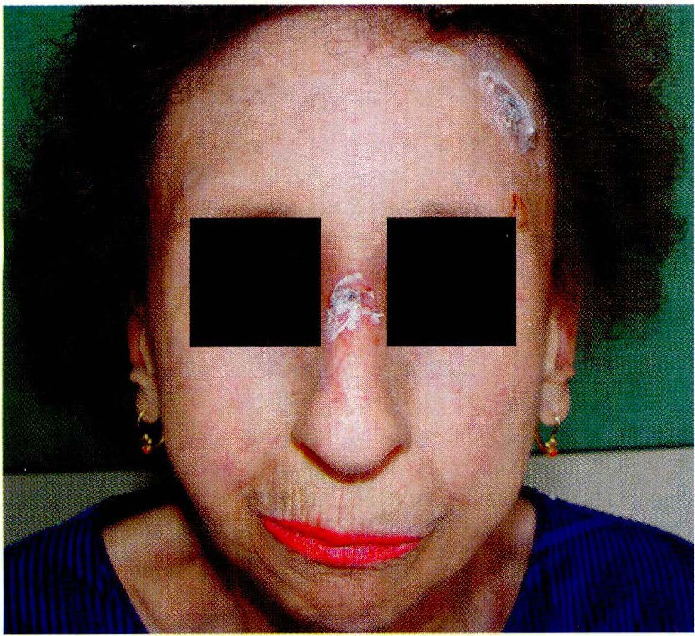
La esclerodermia puede también ocurrir en formas localizadas sin compromiso de órganos internos. Esta variante se conoce como morfea y esclerodermia lineal.
Author Biographies
Ana María Aristizábal Dávila, Instituto de Ciencias de la Salud, CES
RIII Dermatología, Instituto de Ciencias de la Salud (CES), Medellín.
María Adelaida Echeverri Montaño, Instituto de Ciencias de la Salud, CES
RIII Dermatología, CES, Medellín.
Francisco Vargas Carvajal, Universidad Pontificia Bolivariana
RIII Medicina Interna, Universidad Pontificia Bolivariana (UPB), Medellín.
José Fernando Molina Restrepo, Universidad Pontificia Bolivariana
Profesor de Reumatología, UPB, Reumató!ogo Hospital Pablo Tobón Uribe (HPTU), Medel!ín.
Luis Fernando Pinto Peñaranda, Universidad Pontificia Bolivariana
Profesor de Reumatología, UPB, Reumatólogo HPTU, Medellín.
Ángela Zuluaga, Instituto de Ciencias de la Salud, CES
Dermatóloga, Jefe departamento de Dermatología CES, Medellín, Colombia
References
2. Gilliland B. Systemic sclerosis (scleroderma). En: Kasper D, Braunwald E, Fauci A, Dan Longo H, Jameson J. Harrison' s Principies of Interna! Medicine. 1937-45.
3. Renaudineau Y, Revelen R, Levy Y. Anti endhotelial cell antibodies in systemic sclerosis. Clin Diagn Lab lmmunol. 1999; 6:156-6.
4. Ho KT, Reveille JO. The clinical relevance of auto antibodies in scleroderma. Arthritis Res Ther. 2003;5:80-93.
https://doi.org/10.1186/ar628
5. Johnson RW, Tew MB, Arnett Fe. The genetics of systemic sclerosis. Curr Rheumatol Rep. 2002;4:99-107.
https://doi.org/10.1007/s11926-002-0004-2
6. Mayes MD. Epidemiologic studies of environmental agents and systemic autoimmune diseases. Enviran Health Perspect. 1999; 107(suppl5):743-8.
https://doi.org/10.1289/ehp.99107s5743
7. Arlett CM, Smith JB, Jimenez SA. ldentification of fetal DNA and cells in skin lesions from women with systemic sclerosis. N Engl J Med. 1998;338: 1186-91.
https://doi.org/10.1056/NEJM199804233381704
8. Nelson JL. Microchimerism and scleroderma: an update. Curr Rheumatol Rep. 2003;5:154-9
https://doi.org/10.1007/s11926-003-0044-2
9. Burastero SE,Galbiatis, Vassallo A, et al Cellular Microchimerism as a lifelong physiologic status in parous women: an immunologic basis for its amplification in patients with systemic sclerosis. Arhtritis Rheumat. 2003;48: 1109-16.
https://doi.org/10.1002/art.10888
10. Hitraya EG, Varga J, Arlett CM,Jimenez SA. ldentifica tion of elements in the promoter of the a1 (1) procollagen gene involved in its upregulated expression in systemic sclerosis. Arhtritis Rheumat. 1998;41 :2048-58.
https://doi.org/10.1002/1529-0131(199811)41:11<2048::AID-ART21>3.0.CO;2-X
11. Le Roy EC. Systemic sclerosis. A vascular perspective. Rheum Dis Clin North Am. 1996;22:695-708.
https://doi.org/10.1016/S0889-857X(05)70296-9
12. Herrick AL. Vascular function in systemic sclerosis. Curr Opin Rheum. 2000; 12:527-33.
https://doi.org/10.1097/00002281-200011000-00009
13. Xu S, Denton CP, Holmes A, Dashwood Mr, et al. Endothelins: effect on matrix biosynthesis and proliferation in normal and scleroderma fibroblast. J Cardiovasc Pharmacol. 1998;suppl 1 :s 360-3.
14. Mayes MD. Classification and Epidemiology of scleroderma . Seminars in cutaneous Medicine and Surgery. 1998;7(1 ):22-6.
https://doi.org/10.1016/S1085-5629(98)80058-8
15. Flavahan NA, Flavahan S, Mitra S. The vasculopathy of Raynaud's phenomenon and scleroderma. Rheum Dis Clin North Am. 2003; 29:275-91.
https://doi.org/10.1016/S0889-857X(03)00021-8
16. Cutolo M, Grassi W, Matucci M. Raynaud's phenomenon and the role of capillaroscopy . Arhtritis Rheumat. 2003;48:23-30.
https://doi.org/10.1002/art.11310
17. Rodnan GP, Lipinski E, Luksick J. Skin thickness and collagen content in progressive systemic sclerosis and localized scleroderma Arthritis Rheumat. 1979;22: 130-40.
https://doi.org/10.1002/art.1780220205
18. Lovell CR, Jayson MI. Joint involvement in systemic sclerosis. Scand J Rheumatol. 1979; 8: 154-60.
https://doi.org/10.3109/03009747909114448
19. Coghlan JG, Mukerjee D. The heart and pulmonary vasculature in scleroderma clinical features and pathobiology. Curr Opin Rheum. 2001; 13:495-9.
https://doi.org/10.1097/00002281-200111000-00008
20. Steen W. Scleroderma renal crisis. Rheum Dis Clin North Am. 2003;29:315-33.
https://doi.org/10.1016/S0889-857X(03)00016-4
21. Reveille J. Ethnicity and race and systemic sclerosis: how it affects susceptibility, severity, antibody genetics and clinical manifestations. Curr Rheumatol Rep. 2003;5: 160-67.
https://doi.org/10.1007/s11926-003-0045-1
22. Londoño JC. Escleroderma. Estudio descriptivo de 102 pacientes en el Hospital San Juan de Dios , Santa Fé de Bogota. Rev Col de Reumat. 1998.5(3):131-41.
23. Lin AT, Clements PJ, Furst DE. Update on diseasemodifying antirheumatic drugs in the treatment of systemic sclerosis. Rheum Dis Clin North Am. 2003;29:409-26.
https://doi.org/10.1016/S0889-857X(03)00026-7
24. Herrick A. Treatment of Raynaud's phenomenon: new insights and developments. Curr Rheumatol Rep. 2003;5:168-74.
https://doi.org/10.1007/s11926-003-0046-0
25. Wemple MA, Raugi G. CREST Syndrome. www.emedicine.com. 2001
26. Akesso A, Wolleheim FA. Organ manifestations in 100 patients with progressive systemic sclerosis: a comparison between the CREST syndrome and diffuse scleroderma. Br J Rheumat. 1989;28(4):281-6.
https://doi.org/10.1093/rheumatology/28.4.281
27. Hazen PG, Walker AE, Carney J.F et al. Cutaneous calcinosis of scleroderma. Successful treatment with intralesional adrenal steroids. Arch Dermatol.1982; 118(5):366-7.
https://doi.org/10.1001/archderm.1982.01650170080035
28. Dent CE, Stamp TC. Treatment of calcinosis circumscripta with probenecid. Br Med J. 1972; Jan 22;1 (794):216-8.
https://doi.org/10.1136/bmj.1.5794.216
29. Farah MJ, Palmieri GM, Sebes JL et al: The effect of diltiazem on calcinosis in a patient with CREST syndrome. Arhtritis Rheumat. 1990;33(8):1287-90.
https://doi.org/10.1002/art.1780330834
30. Berger RG, Featherstone GL, et al . Treatment of calcinosis universalis with low dose warfarin. Am J Med. 1987;83(1 ):72-6.
https://doi.org/10.1016/0002-9343(87)90499-2
31. Vierra E, Bari B. Morphea and localized scleroderma in children. Seminars in cutaneous Medicine and Surgery. 1999;18(3):210-25.
https://doi.org/10.1016/S1085-5629(99)80019-4
32. Tu J, Eisen A. Scleroderma. En: Freedberg IM, Eisen A, Wolf K, et al Fitzpatrick's Dermatology in General Medicine. 1999; 2023-33.
33. Rowell NR, Goodfield MJ. The connective tissue diseases. En: Champion R.H, Burton J.L, Burns O.A, et al. Rook / Wilkinson/ Ebling Textbook of Dermatolo·gy.1998; 2501-44.
34. Cunnignhan BB, Landells ID, Langman C et al. Topical calcipotriene for morphea/ linear scleroderma. J Am Acad Dermatol. 1998; 39: 211-15.
https://doi.org/10.1016/S0190-9622(98)70077-5
35. Seyger MMB, Van Den Hoogen FHJ, de Boo T et al. Low dose methotrexate in the treatment of widespread morphea. J Am Acad Dermatol. 1998; (39)220-25.
https://doi.org/10.1016/S0190-9622(98)70079-9
36. Joly P, Bamberg N, Crickx B et al. Treatment of severe forms of localized scleroderma with oral corticosteroids: Follow-up study on 17 patients. Arch Dermatol. 1994; 130: 663-64.
https://doi.org/10.1001/archderm.1994.01690050133027
How to Cite
Downloads
Downloads
Published
How to Cite
Issue
Section

| Article metrics | |
|---|---|
| Abstract views | |
| Galley vies | |
| PDF Views | |
| HTML views | |
| Other views | |


