
Cutaneus wound healing.
Keywords:
wound, scar, inflammation, cell proliferationAbstract
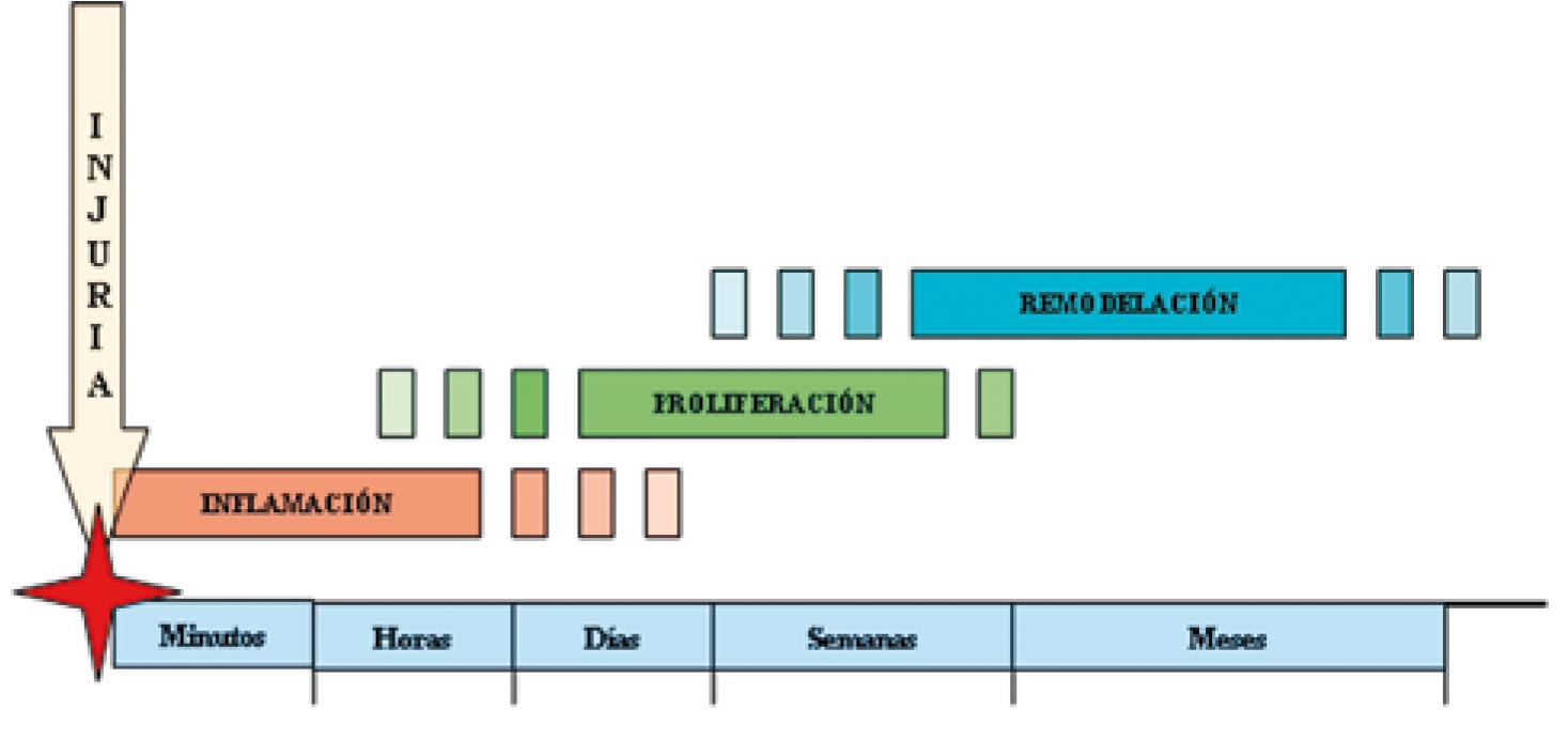
The mechanisms of wound healing come into function after an injury that alters the skin surface continuity. Three phases have been identified in the process: inflammatory, proliferative and tissue remodeling. In the inflammatory phase there is local cell infiltration, release of compounds transported by blood and activation of the coagulation system. In the proliferative phase there is formation of new tissue, due to cell growth and migration, and the participation of diverse adherence molecules. Tissue remodeling corresponds to the last phase, in which a stable tissue develops, similar to the one previously present before the injury, known as scar. The participation of growth factors, cytokines and diverse blood components are fundamental for the functional restoration of the affected area.
Author Biography
Joaquin Benavides
Residente de Dermatología. Universidad del Valle, Cali, Colombia.
References
2. Baum C, Arpey C. Normal Cutaneous Wound Healing: Clinical Correlation with Cellular and Molecular Events. Dermatology surgery 2005; 31:674-86.
3. Grose W, Grose S, Grose R. Regulation of Wound Healing by Growth Factors and Cytokines. Physiol Rev. Am J Pathol 2003; 83: 835-70.
4. Laurens N, Koolwijk P, De Maat M.P.M. Fibrin structure and wound healing. J Thromb Haemost 2006; 4: 932-9.
5. Roh c, Lyle S. Cutaneous stem cell and Wound Healing. Pediatric Research 2006; 59:100R-103R.
6. Frank S, Kämpfer H, Wetzeler C, et al. Nitric oxide drives skin repair: Novel functions of an established mediador. Kidney International 2002; 61:882-8.
7. Schwartz J.R, Marsh R.G, Draelos Z.D. Zinc and Skin Health: Overview of Physiology and Pharmacology. Dermatol Surg 2005; 31:837-47.
8. Scheid A, Meuli M, Gassmann M et al. Genetically modified mouse models in studies on cutaneous wound healing. Exp Physiol 2000; 85:687-704.
9. Otrock Z, Mahfouz R, Makarem J et al. Understanding the biology of angiogenesis: Review of the most important molecular mechanisms. Blood cell, Molecules, and Diseases 2007;39:212-220.
10. Agaiby A, Dyson M. Immuno-infammatory cell dynamics during cutaneous wound healing. J. Anat 1999; 195: 531-542.
11. Wong T, Mcgrath J.A, Navsaria H. The role of fibroblasts in tissue engineering and regeneration. British Journal of Dermatology 2007; 156:1149-55.
12. Vaughan MB, Howard EW, Tomasek JJ. Transforming growth factor β1 promotes the morphological and functional differentiation of the myofibroblast. Exp Cell Res 2000; 257:180- 9.
13. Becker BF, Heindl B, Kupatt C, et al. Endothelial function and hemostasis. Z Kardiol 2000; 89:160–7.
14. Li J, Zhang Y, Kirsner R. Angiogenesis in wound repair: Angiogenic growth factors and the extracellular matrix. Microsc. Res. Tech. 2003; 60:107-14.
15. Sternlicht M, Werb Z. How matrix metalloproteinases regulate cell behavior. Annu Rev Cell Dev Biol 2001; 17:463–516.
16. Nagase H, Woessner JF Jr. Matrix metalloproteinases. J Biol Chem 1999; 274:21491–4.
17. Gallo RL. Proteoglycans and cutaneous vascular defense and repair. J Invest Dermatol Symp Proc 2000; 5:55–60.
How to Cite
Downloads
Downloads
Published
How to Cite
Issue
Section

| Article metrics | |
|---|---|
| Abstract views | |
| Galley vies | |
| PDF Views | |
| HTML views | |
| Other views | |


