
Quantification of melanocytes in facial skin: another help for evaluation of resection borders of melanoma.
Keywords:
Melanocytes, basocellular carcinoma, melanoma, malign lentigo, melanoma in situAbstract
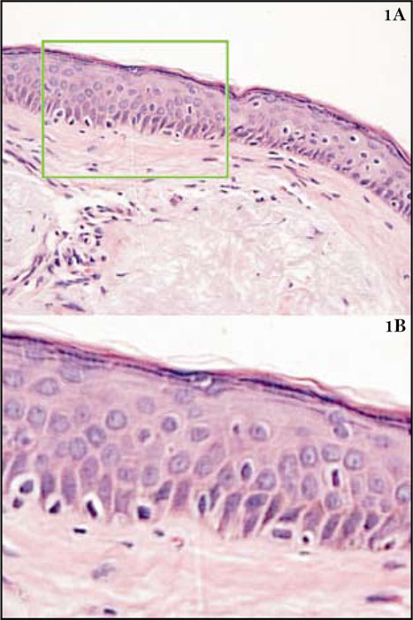
Introduction: Epidermal melanocytes are widely separated from each other, and are surrounded by a hallo; they have a light cytoplasm and picnotic nucleus, smaller than in keratocytes. It is difficult to differenciate facial changes due to solar exposition and a melanoma in situ, likewise to establish if the resection borders of a melanoma in situ have a tumor cells or if the melanocytes there have just changed due to sunlight.
Objective: To quantify the number of melanocytes in normal adults and in resection borders of melanomas without tumor cells in basocellular carcinomas and melanomas in situ in the malar skin.
Material and methods: 25 cheek specimens of 40 year old adults with type I-II skin were studied, seven autopsies of men, eleven of basocellular carcinoma borders and seven of in situ melanoma resection borders free of tumor. Using hematoxylin-eosin staining, three observers counted basal melanocytes by linear millimeter in each specimen, using an Axiophot Zeiss photomicroscope.
Results: In a linear millimeter (three 40X fields), the number of melanocytes was of 18±3 in normal skin, 22±7 in basocellular carcinoma borders and 30±9 in situ melanoma.
Conclusions: the maximum number of melanocytes in a 40X field in the tissues studied should not go over 7.5±4 (30 melanocytes) per linear mm. A larger number alerts the clinician and should be added to other changes to determine if there are persistent melanoma cells in situ.
Author Biographies
Ángel Cruz-Roa
Ingeniero de Sistemas, Grupo de Microscopía y Análisis de Imágenes, Instituto Nacional de Salud, Bogotá, D.C., Colombia.
Ladys Sarmiento
Profesional especializado, Grupo de Microscopía y Análisis de Imágenes, Instituto Nacional de Salud, Bogotá, D.C., Colombia.
María Leonor Caldas
Profesional especializado, Grupo de Microscopía y Análisis de Imágenes, Instituto Nacional de Salud, Bogotá, D.C., Colombia.
Gerzaín Rodríguez
Profesor, Facultad de Medicina, Universidad de la Sabana, Chía, Colombia.
References
2. Cochran A. The incidence of melanocytes in normal human skin. J Invest Dermatol. 1970;55:65-70.
3. Ackerman AB, Cerroni L, Kerl H. Pitfalls in histopathological diagnosis of malignant melanoma. Philadelphia: Lea & Febiger; 1994. 227-9.
4. Weyers W, Bonczkowitz M, Weyers I, Bittinger A, Schill WB. Melanoma in situ versus melanocytic hyperplasia in sundamaged skin. Assessment of the significance of histopathologic criteria for differential diagnosis. Am J Dermatopathol. 1996;18:500-6.
5. Rueda X, Acosta A, Aristizábal L, Fierro E. Guías de práctica clínica para el tratamiento del carcinoma basocelular. Rev Asoc Colomb Dermatol. 2008;16:102-17.
6. Cochran A. Studies on the melanocytes of the epidermis adjacent to tumors. J Invest Dermatol. 1971;57:38-43.
7. Wong CK. A study of melanocytes in the normal skin surrounding malignant melanoma. Dermatologica. 1970;141:215-25.
8. Fallowfield ME, Cook MG. Epidermal melanocytes adjacent to melanoma and the field change effect. Histopathology. 1990;17:397-400.
9. Ackerman AB, Briggs PL, Bravo F. Differential diagnosis in dermatopathology. III. Philadelphia: Lea & Febiger; 1993. 66-7.
How to Cite
Downloads
Downloads
Published
How to Cite
Issue
Section

| Article metrics | |
|---|---|
| Abstract views | |
| Galley vies | |
| PDF Views | |
| HTML views | |
| Other views | |


