
What is your diagnosis?.
Keywords:
carcinoma, basal cell, dermatology, pathologyAbstract
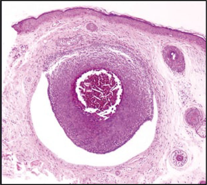
Basal cell carcinoma has two components: epithelial and stromal. The presence of a cleft between these two components in histologic sections is of diagnostic value. The cleft may be so extensive that it leads to the disappearance of the epithelial component displaying only a fibrous stroma with mucinous change, finding that is a clue to diagnose basal cell carcinoma as it is illustrated in this article. This is an essential knowledge for any microscopist that examines skin biopsies as it was emphasized by Ackerman in his clues to diagnosis in dermatopathology.
Author Biographies
Diana Lulú Ruiz
Residente de primer año de dermatología de la Universidad Nacional de Colombia
Ricardo González
Residente de primer año de patología anatómica y clínica de la Universidad Nacional de Colombia
References
1. Ackerman AB, Jacobson M, Vitale P. Clues to diagnosis in dermatopathology I. Chicago: ASCP Press; 1991.
2. Haupt HM, Stern JB, Dilaimy MS. Basal cell carcinoma, clues to its presence in histologic sections when the initial slide is nondiagnostic. Am J Surg Path. 2000;24:1291-4.
3. McArdle JP, Roff BT, Muller HK. Characterization of retraction spaces in basal cell carcinoma using an antibody to type IV collagen. Histopathology. 1984;8:447-55.
4. Kallioinen M, Autio-Harmainen H, Dammert K, Risteli J, Risteli L. Discontinuity of the basement membrane in fibrosing basocellular carcinomas and basosquamous carcinomas of the skin: An inmunohistochemical study with human laminin and type IV collagen antibodies. J Invest Dermatol. 1984;82:248-51.
How to Cite
1.
Ruiz DL, González R. What is your diagnosis?. rev. asoc. colomb. dermatol. cir. dematol. [Internet]. 2019 Feb. 13 [cited 2024 Jul. 22];17(4):245-7. Available from: https://revista.asocolderma.org.co/index.php/asocolderma/article/view/182
Downloads
Download data is not yet available.
Downloads
Published
2019-02-13
How to Cite
1.
Ruiz DL, González R. What is your diagnosis?. rev. asoc. colomb. dermatol. cir. dematol. [Internet]. 2019 Feb. 13 [cited 2024 Jul. 22];17(4):245-7. Available from: https://revista.asocolderma.org.co/index.php/asocolderma/article/view/182
Issue
Section
Make the diagnosis yourself. Part 2

| Article metrics | |
|---|---|
| Abstract views | |
| Galley vies | |
| PDF Views | |
| HTML views | |
| Other views | |


