
Superficial mycosis
DOI:
https://doi.org/10.29176/2590843X.269Keywords:
superficial mycoses, cutaneous mycoses, seborrheic dermatitis, tinea versicolor, tinea nigra, white piedra, black piedraAbstract
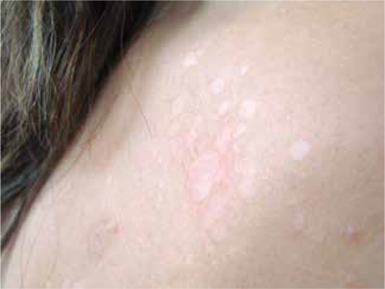
Superficial mycoses are fungal infections involving the stratum corneum and/ or adnexal structures without invading deeper. It includes tinea versicolor, seborrheic dermatitis, tinea nigra, white piedra and black piedra. It produces varied clinical lesions depending on the affected site and causal agent. Generally, it produces little or no immune response so treatment can be performed with topical medications usually and only in severe or refractory cases requiring use of systemic medication.
Author Biographies
Lina Tatiana Acosta
Médica, residente de Dermatología, Universidad CES, Medellín, Colombia
Nora Cardona
Médica dermatóloga, docente de Micología, Universidad CES, Medellín, Colombia
References
1. Sánchez-Saldaña L, Matos-Sánchez R, Kumakawa H. Infecciones micóticas superficiales. Dermatol Peru. 2009;19:226-66.
2. Bonifaz A, Gómez-Daza F, Paredes V, Ponce RM. Tinea versicolor, tinea nigra, white piedra, and black piedra. Clin Dermatol. 2010;28:140-5.
3. Crespo-Erchiga V, Gómez-Moyano E, Crespo M. La pitiriasis versicolor y las levaduras del género Malassezia. Actas DermoSifiliográficas. 2008;99:764-71.
4. Crespo-Erchiga V, Florencio VD. Malassezia yeasts and pityriasis versicolor. Curr Opin Infect Dis. 2006;19:139-47.
5. Padilla-Desgarennes MC. Pitiriasis versicolor. Dermatol Rev Mex. 2005;49:157-67.
6. Gupta AK, Bluhm R, Summerbell R. Pityriasis versicolor. J Eur Acad Dermatol Venereol JEADV. 2002;16:19-33.
7. Schwartz RA. Superficial fungal infections. Lancet. 2004;364:1173-82.
8. Hafez M, el-Shamy S. Genetic susceptibility in pityriasis versicolor. Dermatologica. 1985;171:86-8.
9. Cabañes FJ, Hernández JJ, Castellá G. Molecular analysis of Malassezia sympodialis -related strains from domestic animals. J Clin Microbiol. 2005;43:277-83.
10. Martín-González MT, Crespo-Erchinga V, Samaniego-González E, Gómez-Moyano E. Distribución de las especies de Malassezia en pacientes con pitiriasis versicolor y en individuos sanos. Piel. 2010;25:552-60.
11. Crespo V, Delgado V. Malassezia species in skin diseases. Curr Opin Infect Dis. 2002;15:133-42.
12. Han A, Calcara DA, Stoecker WV, Daly J, Siegel DM, Shell A. Evoked scale sign of tinea versicolor. Arch Dermatol. 2009;145:1078.
13. Vives R, Valcayo A. Tratamiento de la pitiriasis versicolor. Form Médica Contin en Aten Primaria. 2002;9:110-8.
14. Payle B, Serrano L, Bieley HC, Reyes BA. Albert’s solution versus potassium hydroxide solution in the diagnosis of tinea versicolor. Int J Dermatol. 1994;33:182-3.
15. Drake LA, Dinehart SM, Farmer ER, Goltz RW, Graham GF, Hordinsky MK, et al. Guidelines of care for superficial mycotic infections of the skin: Pityriasis (tinea) versicolor. Guidelines/ Outcomes Committee. American Academy of Dermatology. J Am Acad Dermatol. 1996;34:287-9.
16. Meisel C. 10-day therapy of pityriasis versicolor with ketoconazole. Z Für Hautkrankh. 1983;58:1130-6.
17. Fernández-Nava HD, Laya-Cuadra B, Tianco EA. Comparison of single dose 400 mg versus 10-day 200 mg daily dose ketoconazole in the treatment of tinea versicolor. Int J Dermatol. 1997;36:64-6.
18. Sadeque JB, Shahidullah M, Shah OR, Kamal M. Systemic ketoconazole in the treatment of tinea versicolor. Int J Dermatol. 1995;34:504-5.
19. Bhogal CS, Singal A, Baruah MC. Comparative efficacy of ketoconazole and fluconazole in the treatment of pityriasis versicolor: A one year follow-up study. J Dermatol. 2001;28:535-9.
20. Farschian M, Yaghoobi R, Samadi K. Fluconazole versus ketoconazole in the treatment of tinea versicolor. J Dermatol Treat. 2002;13:73-6.
21. Del Rosso JQ, Gupta AK. The use of intermittent itraconazole therapy for superficial mycotic infections: A review and update on the «one week» approach. Int J Dermatol. 1999;38(Suppl.2):28-39.
22. Delescluse J. Itraconazole in tinea versicolor: A review. J Am Acad Dermatol. 1990;23:551-4.
23. Balfour JA, Faulds D. Terbinafine. A review of its pharmacodynamic and pharmacokinetic properties, and therapeutic potential in superficial mycoses. Drugs. 1992;43:259-84.
24. Rausch LJ, Jacobs PH. Tinea versicolor: Treatment and prophylaxis with monthly administration of ketoconazole. Cutis. 1984;34:470-1.
25. Faergemann J, Djärv L. Tinea versicolor: Treatment and prophylaxis with ketoconazole. Cutis. 1982;30:542-50.
26. Faergemann J, Gupta AK, Al Mofadi A, Abanami A, Shareaah AA, Marynissen G. Efficacy of itraconazole in the prophylactic treatment of pityriasis (tinea) versicolor. Arch Dermatol. 2002;138:69-73.
27. Stefanaki I, Katsambas A. Therapeutic update on seborrheic dermatitis. Skin Ther Lett. 2010;15:1-4.
28. Sampaio ALSB, Mameri ACA, Vargas TJ de S, Ramos-e-Silva M, Nunes AP, Carneiro SC da S. Seborrheic dermatitis. An Bras Dermatol. 2011;86:1061-71.
29. Leone R. Presence and significance of Pityrosporon ovale in pityriasis of the scalp, in figured seborrheic eczema and in various squamous dermatoses. Note III. Cultural, biological and biochemical properties of Pityrosporon ovale, with special reference to the affinity for lipidic substances. Minerva Dermatol. 1952;27:123-7.
30. Inamadar AC, Palit A. The genus Malassezia and human disease. Indian J Dermatol Venereol Leprol. 2003;69:265-70.
31. Schechtman RC, Midgley G, Hay RJ. HIV disease and Malassezia yeasts: A quantitative study of patients presenting with seborrhoeic dermatitis. Br J Dermatol. 1995;133:694-8.
32. Bukvic Z, Kralj M, Basta-Juzbašic A, Lakoš I. Seborrheic dermatitis: An update. Acta Dermatovenerol Croat ADC. 2012;20:98-104.
33. Gupta AK, Batra R, Bluhm R, Boekhout T, Dawson TL Jr. Skin diseases associated with Malassezia species. J Am Acad Dermatol. 2004;51:785-98.
34. Nakabayashi A, Sei Y, Guillot J. Identification of Malassezia species isolated from patients with seborrhoeic dermatitis, atopic dermatitis, pityriasis versicolor and normal subjects. Med Mycol Off Publ Int Soc Hum Anim Mycol. 2000;38:337-41.
35. Dawson TL Jr. Malassezia globosa and restricta: Breakthrough understanding of the etiology and treatment of dandruff and seborrheic dermatitis through whole-genome analysis. J Investig Dermatol Symp Proc Soc Investig Dermatol Inc Eur Soc Dermatol Res. 2007;12:15-9.
36. Tajima M, Sugita T, Nishikawa A, Tsuboi R. Molecular analysis of Malassezia microflora in seborrheic dermatitis patients: comparison with other diseases and healthy subjects. J Invest Dermatol. 2008;128:345-51.
37. Gupta AK, Bluhm R. Seborrheic dermatitis. J Eur Acad Dermatol Venereol JEADV. 2004;18:13-26.
38. Ferrándiz C, Ferrándiz-Pulido C. Dermatitis seborreica facial. Aspectos patogénicos y terapéuticos. Piel. 22:393-8.
39. Foley P, Zuo Y, Plunkett A, Merlin K, Marks R. The frequency of common skin conditions in preschool-aged children in Australia: Seborrheic dermatitis and pityriasis capitis (cradle cap). Arch Dermatol. 2003;139:318-22.
40. Poindexter GB, Burkhart CN, Morrell DS. Therapies for pediatric seborrheic dermatitis. Pediatr Ann. 2009;38:333-8.
41. Peyrí J, Lleonart M, Grupo español del Estudio SEBDERM. Clinical and therapeutic profile and quality of life of patients with seborrheic dermatitis. Actas Dermo-Sifiliográficas. 2007;98:476-82.
42. Peter RU, Richarz-Barthauer U. Successful treatment and prophylaxis of scalp seborrhoeic dermatitis and dandruff with 2% ketoconazole shampoo: Results of a multicentre, double-blind, placebo-controlled trial. Br J Dermatol. 1995;132:441-5.
43. Segal R, David M, Ingber A, Lurie R, Sandbank M. Treatment with bifonazole shampoo for seborrhea and seborrheic dermatitis: A randomized, double-blind study. Acta Derm Venereol. 1992;72:454-5.
44. Faergemann J. Seborrhoeic dermatitis and Pityrosporum orbiculare: treatment of seborrhoeic dermatitis of the scalp with miconazole-hydrocortisone (Daktacort), miconazole and hydrocortisone. Br J Dermatol. 1986;114:695-700.
45. Lorette G, Ermosilla V. Clinical efficacy of a new ciclopiroxolamine/zinc pyrithione shampoo in scalp seborrheic dermatitis treatment. Eur J Dermatol EJD. 2006;16:558-64.
46. Schmidt-Rose T, Braren S, Fölster H, Hillemann T, Oltrogge B, Philipp P, et al. Efficacy of a piroctone olamine/climbazol shampoo in comparison with a zinc pyrithione shampoo in subjects with moderate to severe dandruff. Int J Cosmet Sci. 2011;33:276-82.
47. Naldi L, Rebora A. Clinical practice. Seborrheic dermatitis. N Engl J Med. 2009;360:387-96.
48. Ortonne JP, Lacour JP, Vitetta A, Le Fichoux Y. Comparative study of ketoconazole 2% foaming gel and betamethasone dipropionate 0.05% lotion in the treatment of seborrhoeic dermatitis in adults. Dermatol Basel Switz. 1992;184:275-80.
49. Parsad D, Pandhi R, Negi KS, Kumar B. Topical metronidazole in seborrheic dermatitis –a double-blind study. Dermatol Basel Switz. 2001;202:35-7.
50. Koca R, Altinyazar HC, Estürk E. Is topical metronidazole effective in seborrheic dermatitis? A double-blind study. Int J Dermatol. 2003;42:632-5.
51. Cook BA, Warshaw EM. Role of topical calcineurin inhibitors in the treatment of seborrheic dermatitis: A review of pathophysiology, safety, and efficacy. Am J Clin Dermatol. 2009;10:103-18.
52. Warshaw EM, Wohlhuter RJ, Liu A, Zeller SA, Wenner RA, Bowers S, et al. Results of a randomized, double-blind, vehicle-controlled efficacy trial of pimecrolimus cream 1% for the treatment of moderate to severe facial seborrheic dermatitis. J Am Acad Dermatol. 2007;57:257-64.
53. Pirkhammer D, Seeber A, Hönigsmann H, Tanew A. Narrowband ultraviolet B (ATL-01) phototherapy is an effective and safe treatment option for patients with severe seborrhoeic dermatitis. Br J Dermatol. 2000;143:964-8.
54. Ford GP, Farr PM, Ive FA, Shuster S. The response of seborrhoeic dermatitis to ketoconazole. Br J Dermatol. 1984;111:603-7.
55. Morales CA, Sánchez G. Efectividad del ketoconazol oral en el tratamiento de la dermatitis seborreica moderada a grave. Rev Asoc Colomb Dermatol. 2011;19:109-16.
56. Vena GA, Micali G, Santoianni P, Cassano N, Peruzzi E. Oral terbinafine in the treatment of multi-site seborrhoic dermatitis: A multicenter, double-blind placebo-controlled study. Int J Immunopathol Pharmacol. 2005;18:745-53.
57. Cömert A, Bekiroglu N, Gürbüz O, Ergun T. Efficacy of oral fluconazole in the treatment of seborrheic dermatitis: A placebocontrolled study. Am J Clin Dermatol. 2007;8:235-8.
58. Geissler SE, Michelsen S, Plewig G. Very low dose isotretinoin is effective in controlling seborrhea. J Dtsch Dermatol Ges J Ger Soc Dermatol JDDG. 2003;1:952-8.
59. Barzilai A, David M, Trau H, Hodak E. Seborrheic dermatitis-like eruption in patients taking isotretinoin therapy for acne: Retrospective study of five patients. Am J Clin Dermatol. 2008;9:255-61.
60. Babel DE, Pelachyk JM, Hurley JP. Tinea nigra masquerading as acral lentiginous melanoma. J Dermatol Surg Oncol. 1986;12:502-4.
61. McGinnis MR, Schell WA, Carson J. Phaeoannellomyces and the Phaeococcomycetaceae, new dematiaceous blastomycete taxa. Sabouraudia. 1985;23:179-88.
62. Cabrera R, Sabatin N, Urrutia M, Sepúlveda R. Tinea nigra: A allochthonous case report in Chile. Rev Chil Infectol Órgano Of Soc Chil Infectol. 2013;30:90-3.
63. Hughes JR, Moore MK, Pembroke AC. Tinea nigra palmaris. Clin Exp Dermatol. 1993;18:481-2.
64. Pérez C, Colella MT, Olaizola C, Hartung C, Magaldi S, MataEssayag S. Tinea nigra: Report of twelve cases in Venezuela. Mycopathologia. 2005;160:235-8.
65. Muellenhoff M, Cukrowski T, Morgan M, Miller R. Enlarging pigmented patches on the hand. Int J Dermatol. 2003;42:810-1.
66. Cabañes FJ, Bragulat MR, Castellá G. Hortaea werneckii isolated from silicone scuba diving equipment in Spain. Med Mycol Off Publ Int Soc Hum Anim Mycol. 2012;50:852-7.
67. Bonifaz A, Badali H, de Hoog GS, Cruz M, Araiza J, Cruz MA, et al. Tinea nigra by Hortaea werneckii, a report of 22 cases from Mexico. Stud Mycol. 2008;61:77-82.
68. Ritchie EB, Taylor TE. A study of tinea nigra palmaris. Report of a case and inoculation experiments. Arch Dermatol. 1964;89:601-3.
69. Blank H. Tinea nigra: A twenty-year incubation period? J Am Acad Dermatol. 1979;1:49-51.
70. Severo LC, Bassanesi MC, Londero AT. Tinea nigra: Report of four cases observed in Rio Grande do Sul (Brazil) and a review of Brazilian literature. Mycopathologia. 1994;126:157-62.
71. Vanvelsor H, Singletary H. Tinea nigra palmaris. A report of 15 cases from coastal North Carolina. Arch Dermatol. 1964;90:59-61.
72. Tseng SS, Whittier S, Miller SR, Zalar GL. Bilateral tinea nigra plantaris and tinea nigra plantaris mimicking melanoma. Cutis Cutan Med Pract. 1999;64:265-8.
73. Gupta G, Burden AD, Shankland GS, Fallowfield ME, Richardson MD. Tinea nigra secondary to Exophiala werneckii responding to itraconazole. Br J Dermatol. 1997;137:483-4.
74. Piliouras P, Allison S, Rosendahl C, Buettner PG, Weedon D. Dermoscopy improves diagnosis of tinea nigra: A study of 50 cases: Dermoscopy improves tinea nigra diagnosis. Australas J Dermatol. 2011;52:191-4.
75. Criado PR, Delgado L, Pereira GA. Dermoscopy revealing a case of tinea nigra. An Bras Dermatol. 2013;88:128-9.
76. Maldonado I, Fernández L, Leitner R, Vitale RG. Tinea nigra palmaris: A clinical case in Argentina. Rev Argent Microbiol. 2007;39:218-20.
77. Marks JG Jr, King RD, Davis BM. Treatment of tinea nigra palmaris with miconazole. Arch Dermatol. 1980;116:321-2.
78. Burke WA. Tinea nigra: Treatment with topical ketoconazole. Cutis Cutan Med Pract. 1993;52:209-11.
79. Uezato H, Gushi M, Hagiwara K, Kayo S, Hosokawa A, Nonaka S. A case of tinea nigra palmaris in Okinawa, Japan. J Dermatol. 2006;33:23-9.
80. Shannon PL, Ramos-Caro FA, Cosgrove BF, Flowers FP. Treatment of tinea nigra with terbinafine. Cutis Cutan Med Pract. 1999;64:199-201.
81. Rossetto AL, Cruz RCB. Tinea nigra: Successful treatment with topical butenafine. An Bras Dermatol. 2012;87:939-41.
82. Martínez E, Tejada D, Koris Y, Schlager H, Arenas R. Piedra blanca y efluvio telógeno. Una rara asociación. Rev Médica Honduras. 2012;80:58-60.
83. Romero M, Castillo A, Arenas R, Fernández R. Piedra blanca. Revisión de los casos mexicanos y estudio de prevalencia y factores de riesgo de cien pacientes atendidas en la consulta externa de dermatología del Hospital General de Acapulco, Guerrero. Dermatol Rev Mex. 2011;55:3-8.
84. Kalter DC, Tschen JA, Cernoch PL, McBride ME, Sperber J, Bruce S, et al. Genital white piedra: Epidemiology, microbiology, and therapy. J Am Acad Dermatol. 1986;14:982-93.
85. Kiken DA, Sekaran A, Antaya RJ, Davis A, Imaeda S, Silverberg NB. White piedra in children. J Am Acad Dermatol. 2006;55:956-61.
86. Stenderup A, Schønheyder H, Ebbesen P, Melbye M. White piedra and Trichosporon beigelii carriage in homosexual men. J Med Vet Mycol Bi-Mon Publ Int Soc Hum Anim Mycol. 1986;24:401-6.
87. Avram A, Buot G, Binet O, Gracia AM, Cesarini JP. Clinical and mycological study of 11 cases of genitopubic trichosporosis nodosa (white piedra). Ann Dermatol Vénéréologie. 1987;114:819-27.
88. Ruiz-Orozco IM, Hernández-Arana MS, Quiñones-Venegas R, Mayorga J. Piedra blanca. Presentación de tres casos. Piel. 2004;19:239-41.
89. Magalhães AR, Mondino SSB de, Silva M da, Nishikawa MM. Morphological and biochemical characterization of the aetiological agents of white piedra. Mem Inst Oswaldo Cruz. 2008;103:786-90.
90. Pontes ZBV da S, Ramos AL, Lima E de O, Guerra M de F de L, Oliveira NMC, Santos JP dos. Clinical and mycological study of scalp white piedra in the State of Paraíba, Brazil. Mem Inst Oswaldo Cruz. 2002;97:747-50.
91. Viswanath V, Kriplani D, Miskeen AK, Patel B, Torsekar RG. White piedra of scalp hair by Trichosporon inkin. Indian J Dermatol Venereol Leprol. 2011;77:591-3.
92. Tambe SA, Dhurat SR, Kumar CA, Thakare P, Lade N, Jerajani H, et al. Two cases of scalp white piedra caused by Trichosporon ovoides. Indian J Dermatol Venereol Leprol. 2009;75:293-5.
93. Khandpur S, Reddy BS. Itraconazole therapy for white piedra affecting scalp hair. J Am Acad Dermatol. 2002;47:415-8.
94. Guého E, Smith MT, de Hoog GS, Billon-Grand G, Christen R, Batenburg-van der Vegte WH. Contributions to a revision of the genus Trichosporon. Antonie Van Leeuwenhoek. 1992;61:289-316.
95. Guého E, Improvisi L, de Hoog GS, Dupont B. Trichosporon on humans: A practical account. Mycoses. 1994;3710.
96. Shivaprakash MR, Singh G, Gupta P, Dhaliwal M, Kanwar AJ, Chakrabarti A. Extensive white piedra of the scalp caused by Trichosporon inkin: A case report and review of literature. Mycopathologia. 2011;172:481-6.
97. McBride ME, Ellner KM, Black HS, Clarridge JE, Wolf JE. A new Brevibacterium sp. isolated from infected genital hair of patients with white piedra. J Med Microbiol. 1993;39:255-61.
98. Thérizol-Ferly M, Kombila M, Gómez M, Douchet C, Salaun Y, Barrabes A, et al. White piedra and Trichosporon species in equatorial Africa. II. Clinical and mycological associations: An analysis of 449 superficial inguinal specimens. Mycoses. 1994;37:255-60.
99. De La Cruz S, Cortez F, Pereda O, Aleman I. Piedra blanca en pelo de cuero cabelludo. Folia Dermatol Perú. 2008;19:134-7.
100. Roshan A, Janaki C, Parveen B. White piedra in a mother and daughter. Int J Trichology. 2009;1:140.
101. Ríos X, Rojas RF, Hincapié ML. Ocho casos de piedra blanca en población pediátrica. Rev Asoc Colomb Dermatol. 2012;20:175-80.
102. Kanitakis J, Persat F, Piens MA, Picot S, Claudy A. Black piedra: Report of a French case associated with Trichosporon asahii. Int J Dermatol. 2006;45:1258-60.
103. Fischman O. Black piedra in Brazil. A contribution to its study in Manaus (State of Amazonas). Mycopathol Mycol Appl. 1965;25:201-4.
104. Fischman O. Black piedra among Brazilian Indians. Rev Inst Med Trop São Paulo. 1973;15:103-6.
105. Coimbra Júnior CE, Santos RV. Black piedra among the Zoró Indians from Amazônia (Brazil). Mycopathologia. 1989;107:57-60.
106. Figueras MJ, Guarro J, Zaror L. New findings in black piedra infection. Br J Dermatol. 1996;135:157-8.
107. Gip L. Black piedra: The first case treated with terbinafine (Lamisil). Br J Dermatol. 1994;130(Suppl.43):26-8.
How to Cite
1.
Acosta LT, Cardona N. Superficial mycosis. rev. asoc. colomb. dermatol. cir. dematol. [Internet]. 2014 Oct. 1 [cited 2024 Jul. 22];22(4):278-90. Available from: https://revista.asocolderma.org.co/index.php/asocolderma/article/view/269
Downloads
Download data is not yet available.
Downloads
Published
2014-10-01
How to Cite
1.
Acosta LT, Cardona N. Superficial mycosis. rev. asoc. colomb. dermatol. cir. dematol. [Internet]. 2014 Oct. 1 [cited 2024 Jul. 22];22(4):278-90. Available from: https://revista.asocolderma.org.co/index.php/asocolderma/article/view/269
Issue
Section
Review Article

| Article metrics | |
|---|---|
| Abstract views | |
| Galley vies | |
| PDF Views | |
| HTML views | |
| Other views | |


