
Tranverse versus vertical sections in diagnosis of alopecia.
Keywords:
Hair follicle, scalp, biopsy specimens, alopeciaAbstract
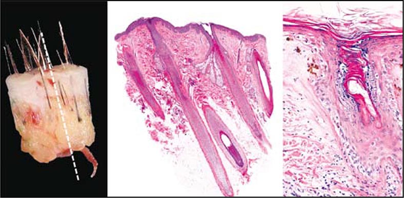
A brief assessment is done over different types of cuts currently done for the study of scalp biopsy specimens (transversal and vertical), analyzing advantages and disadvantages of each one. The current tendency is to combine them whenever possible. In case of having only one biopsy, detail clinic records are to be taken into account to choose the most suitable sample.
Author Biography
Rodrigo Restrepo
Instructor asociado de patología y dermatopatología. Servicio de dermatología, Facultad de Medicina, Universidad Pontificia Bolivariana. Director laboratorio de patología, Clínica Medellín. Medellín, Colombia.
References
1. Mirmirani P, Willey A, Headington JT, et al. Primary cicatricial alopecia: histopathologic findings do not distinguish clinical variants. J Am Acad Dermatol. 2005; 52:637-43.
2. Wain EM, Stefanato CM. Four millimetres: a variable measurement? Br J Dermatol. 2007; 156:404.
3. Elston DM, Ferringer T, Dalton S, Fillman E, Tyler W. A comparison of vertical versus transverse sections in the evaluation of alopecia biopsy specimens. J Am Acad Dermatol. 2005; 53:267-72.
4. Flotte TJ. Transverse sectioning of the scalp (Headington technique) in the 19th century. J Cutan Pathol. 2008; 35:82-5.
5. Headington JT. Transverse microscopic anatomy of the human scalp. A basis for a morphometric approach to disorders of the hair follicle. Arch Dermatol. 1984; 120:449-56.
6. Templeton SF, Santa Cruz DJ, Solomon AR. Alopecia: histologic diagnosis by transverse sections. Semin Diagn Pathol. 1996; 13:2-18.
7. Solomon AR. The transversely sectioned scalp biopsy specimen: the technique and an algorithm for its use in the diagnosis of alopecia. Adv Dermatol. 1994; 9:127-57; discussion 158.
8. Frishberg DP, Sperling LC, Guthrie VM. Transverse scalp sections: a proposed method for laboratory processing. J Am Acad Dermatol. 1996; 35(2 Pt 1):220-2.
9. Sinclair R, Jolley D, Mallari R, Magee J. The reliability of horizontally sectioned scalp biopsies in the diagnosis of chronic diffuse telogen hair loss in women. J Am Acad Dermatol. 2004; 51:189-99.
10. Whiting DA. Diagnostic and predictive value of horizontal sections of scalp biopsy specimens in male pattern androgenetic alopecia. J Am Acad Dermatol. 1993; 28(5 Pt 1):755-63.
11. Shum DT, Lui H, Martinka M, et al. Computerized morphometry and three-dimensional image reconstruction in the evaluation of scalp biopsy from patients with non-cicatricial alopecias. Br J Dermatol. 2003; 148:272-8.
12. Lee MS, Kossard S, Wilkinson B, Doyle JA. Quantification of hair follicle parameters using computer image analysis: a comparison of androgenetic alopecia with normal scalp biopsies.Australas J Dermatol. 1995; 36:143-7.
13. Whiting DA. Histopathology of alopecia areata in horizontal sections of scalp biopsies. J Invest Dermatol. 1995; 104(5 Suppl):26S-27S.
14. Bosseila M, Saad B. Quantitative morphometric analysis of hair follicles in alopecia areata J Dermatol Sci. 2006;44:59-61.
15. Whiting DA, Waldstreicher J, Sanchez M, Kaufman KD. Measuring reversal of hair miniaturization in androgenetic alopecia by follicular counts in horizontal sections of serial scalp biopsies: results of finasteride 1 mg treatment of men and postmenopausal women. J Investig Dermatol Symp Proc. 1999; 4:282-4.
16. Sperling LC. An Atlas of Hair Pathology with Clinical Correlations. Parthenon Publishing: New York; 2003:19.
17. Olsen EA, Bergfeld WF, Cotsarelis G, et al. Workshop on Cicatricial Alopecia. Summary of North American Hair Research Society (NAHRS)-sponsored Workshop on Cicatricial Alopecia, Duke University Medical Center, February 10 and 11, 2001. J Am Acad Dermatol. 2003;48: 103–10.
18. Böer A, Hoene K. Transverse sections for diagnosis of alopecia? Am J Dermatopathol. 2005; 27: 348-52.
19. Elston DM, McCollough ML, Angeloni VL. Vertical and transverse sections of alopecia biopsy specimens: combining the two to maximize diagnostic yield. J Am Acad Dermatol. 1995; 32:454-7.
20. Garcia C, Poletti E. Scalp biopsy specimens: transverse vs vertical sections. Arch Dermatol. 2007; 143:268.
How to Cite
1.
Restrepo R. Tranverse versus vertical sections in diagnosis of alopecia. rev. asoc. colomb. dermatol. cir. dematol. [Internet]. 2019 Feb. 4 [cited 2024 Jul. 22];16(1):23-8. Available from: https://revista.asocolderma.org.co/index.php/asocolderma/article/view/92
Downloads
Download data is not yet available.
Downloads
Published
2019-02-04
How to Cite
1.
Restrepo R. Tranverse versus vertical sections in diagnosis of alopecia. rev. asoc. colomb. dermatol. cir. dematol. [Internet]. 2019 Feb. 4 [cited 2024 Jul. 22];16(1):23-8. Available from: https://revista.asocolderma.org.co/index.php/asocolderma/article/view/92
Issue
Section
Review Article

| Article metrics | |
|---|---|
| Abstract views | |
| Galley vies | |
| PDF Views | |
| HTML views | |
| Other views | |


