
Reparación de heridas cutáneas
Palabras clave:
herida, cicatriz, inflamación, proliferación celularResumen
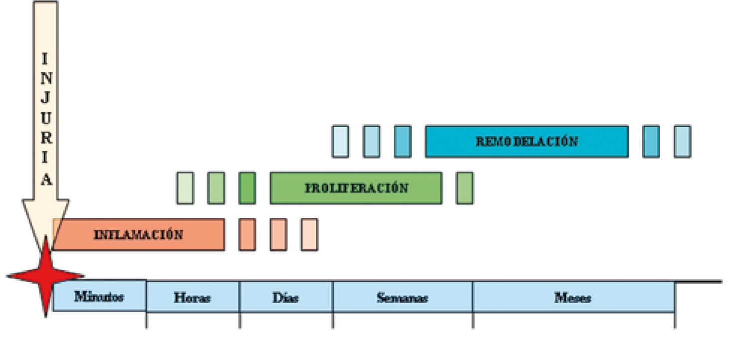
Los mecanismos de reparación de heridas cutáneas se ponen en funcionamiento tras una lesión que altere la continuidad de la superficie. En el proceso se han identificado tres fases: la inflamatoria, la proliferativa y la de remodelación tisular. En la fase inflamatoria hay liberación local de células y compuestos transportados por la sangre y la activación del sistema de coagulación. En la proliferativa hay formación de tejido nuevo, gracias al crecimiento y migración celular y la participación de diversas proteínas de adherencia. La remodelación tisular corresponde a la última fase, cuando se desarrolla un tejido estable, similar al existente previo a la lesión, conocido como cicatriz. La participación de factores de crecimiento, citoquinas y diversos componentes sanguíneos es fundamental para la restauración funcional del área afectada.
Biografía del autor/a
Joaquin Benavides
Residente de Dermatología. Universidad del Valle, Cali, Colombia.
Referencias bibliográficas
2. Baum C, Arpey C. Normal Cutaneous Wound Healing: Clinical Correlation with Cellular and Molecular Events. Dermatology surgery 2005; 31:674-86.
3. Grose W, Grose S, Grose R. Regulation of Wound Healing by Growth Factors and Cytokines. Physiol Rev. Am J Pathol 2003; 83: 835-70.
4. Laurens N, Koolwijk P, De Maat M.P.M. Fibrin structure and wound healing. J Thromb Haemost 2006; 4: 932-9.
5. Roh c, Lyle S. Cutaneous stem cell and Wound Healing. Pediatric Research 2006; 59:100R-103R.
6. Frank S, Kämpfer H, Wetzeler C, et al. Nitric oxide drives skin repair: Novel functions of an established mediador. Kidney International 2002; 61:882-8.
7. Schwartz J.R, Marsh R.G, Draelos Z.D. Zinc and Skin Health: Overview of Physiology and Pharmacology. Dermatol Surg 2005; 31:837-47.
8. Scheid A, Meuli M, Gassmann M et al. Genetically modified mouse models in studies on cutaneous wound healing. Exp Physiol 2000; 85:687-704.
9. Otrock Z, Mahfouz R, Makarem J et al. Understanding the biology of angiogenesis: Review of the most important molecular mechanisms. Blood cell, Molecules, and Diseases 2007;39:212-220.
10. Agaiby A, Dyson M. Immuno-infammatory cell dynamics during cutaneous wound healing. J. Anat 1999; 195: 531-542.
11. Wong T, Mcgrath J.A, Navsaria H. The role of fibroblasts in tissue engineering and regeneration. British Journal of Dermatology 2007; 156:1149-55.
12. Vaughan MB, Howard EW, Tomasek JJ. Transforming growth factor β1 promotes the morphological and functional differentiation of the myofibroblast. Exp Cell Res 2000; 257:180- 9.
13. Becker BF, Heindl B, Kupatt C, et al. Endothelial function and hemostasis. Z Kardiol 2000; 89:160–7.
14. Li J, Zhang Y, Kirsner R. Angiogenesis in wound repair: Angiogenic growth factors and the extracellular matrix. Microsc. Res. Tech. 2003; 60:107-14.
15. Sternlicht M, Werb Z. How matrix metalloproteinases regulate cell behavior. Annu Rev Cell Dev Biol 2001; 17:463–516.
16. Nagase H, Woessner JF Jr. Matrix metalloproteinases. J Biol Chem 1999; 274:21491–4.
17. Gallo RL. Proteoglycans and cutaneous vascular defense and repair. J Invest Dermatol Symp Proc 2000; 5:55–60.
Cómo citar
Descargas
Descargas
Publicado
Cómo citar
Número
Sección

| Estadísticas de artículo | |
|---|---|
| Vistas de resúmenes | |
| Vistas de PDF | |
| Descargas de PDF | |
| Vistas de HTML | |
| Otras vistas | |


