
Reed's nevus.
Keywords:
reed's nevus, biopsy, histologyAbstract
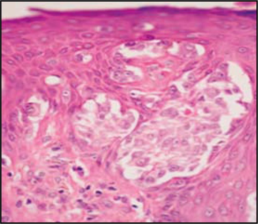
A case of 26 years old patient, with hyperpigmented nodule on her left leg. An excisional biopsy was reported pigmented cell nevus (reed nevus).I will discuss the difficulties involved in evaluating on histopathology.
Author Biography
Mariam Rolón
Dermatopatóloga del Instituto Nacional de Cancerología.
References
1. Reed RJ, Ichinose H, Clark WH Jr, Mihm MC Jr. Common and Uncommon melanocytic nevi and borderline melanomas. Semin Oncol.1975; 2: 119-47.
2. Sagebiel RW, Chinn EK, Egbert BM. Pigmented spindle cell nevus.Clinical and histologic review of 90 cases. Am J Surg Pathol. 1984; 8:645-53.
3. Requena L, Sanchez Yus E. Pigmented spindle cell Nevus. Br J Dermatol.1990; 123:757-63.
4. Sau P, Graham JH, Helwig EB.Pigmented spindle cell nevus: a clinicopathologic analysis of ninety-five cases. J Acad Dermatol.1993;28: 565-71.
5. Smith NP. The pigmented spindle cell tumor of Reed-an underrecognised lesion. Br J Dermatol 1983;109: 39-49.
6. Barnhill RL. Malignant melanoma, dysplastic melanocytic nevi, and Spitz tumors. Histologic classification and characteristics. Clin Plast Surg. 2000; 27:331-60.
How to Cite
1.
Rolón M. Reed’s nevus. rev. asoc. colomb. dermatol. cir. dematol. [Internet]. 2019 Feb. 6 [cited 2024 Jul. 3];16(1):53-4. Available from: https://revista.asocolderma.org.co/index.php/asocolderma/article/view/103
Downloads
Download data is not yet available.
Downloads
Published
2019-02-06
How to Cite
1.
Rolón M. Reed’s nevus. rev. asoc. colomb. dermatol. cir. dematol. [Internet]. 2019 Feb. 6 [cited 2024 Jul. 3];16(1):53-4. Available from: https://revista.asocolderma.org.co/index.php/asocolderma/article/view/103
Issue
Section
Case Report

| Article metrics | |
|---|---|
| Abstract views | |
| Galley vies | |
| PDF Views | |
| HTML views | |
| Other views | |


