
Atypical acral melanocytic hyperplasia vs acral melanoma in situ.
Keywords:
melanoma, pathology, melanocyteAbstract
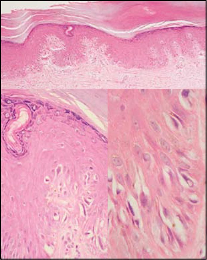
A case of 41 years-old woman with a hiperpigmented macule on her third interdigital right foot with progresive growth. A excisional biopsy was reported as acral melanoma in situ.This article identifies the histopathology features to differential diagnosis.
Author Biography
Mariam Rolón
Dermatopatóloga del Instituto Nacional de Cancerología.
References
1. Kuchelmeister C, Schaumburg-Lever G, Garbe C. Acral cutaneous melanoma in Caucasians. Br J Dermatol. Aug; 143:275- 80.
2. Saida T. Malignant melanoma in situ on the sole of the foot. Its clinical and histopathologic characteristics. Am J Dermatopathol. 1989; 11: 124-30.
3. Saida T. Malignant melanoma on the sole: how to detect the early lesions efficiently. Pigment cell Res 13 suppl 8: 135-139, 2000.
4. Boyd AS, Rapini RP.Acral melanocytic neoplasms:a histolgic analyisisof 158 lesions. J Am Acad Dermatol.1994; 31 (5pt 1):740-5.
5. Signoretti S, Annessi G, Puddu P, Faraggiana T. Melanocytic nevi of palms and soles: a histologycal study according to the plane of section. Am J Surg Pathol. 1999; 23: 283-7.
How to Cite
1.
Rolón M. Atypical acral melanocytic hyperplasia vs acral melanoma in situ. rev. asoc. colomb. dermatol. cir. dematol. [Internet]. 2019 Feb. 5 [cited 2024 Jul. 3];16(1):47-8. Available from: https://revista.asocolderma.org.co/index.php/asocolderma/article/view/99
Downloads
Download data is not yet available.
Downloads
Published
2019-02-05
How to Cite
1.
Rolón M. Atypical acral melanocytic hyperplasia vs acral melanoma in situ. rev. asoc. colomb. dermatol. cir. dematol. [Internet]. 2019 Feb. 5 [cited 2024 Jul. 3];16(1):47-8. Available from: https://revista.asocolderma.org.co/index.php/asocolderma/article/view/99
Issue
Section
Case Report

| Article metrics | |
|---|---|
| Abstract views | |
| Galley vies | |
| PDF Views | |
| HTML views | |
| Other views | |


