
Rhabdomyomatous mesenchymal hamartoma
Keywords:
Hamartoma, pyloric stenosis, hypertrophic, dermisAbstract
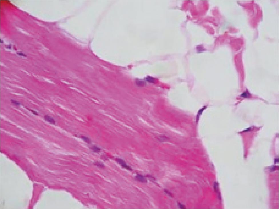
We present a case of a 12-year-old girl with a nodular lesion on her chin, it appeared 6 months previously. Histopathology examination revealed the presence skeletal muscle fibers in reticular dermis and subcutaneous tissues associated with normal-appearing mesenchymal elements. She had a diagnosis of infantile hypertrophic pyloric stenosis when she was born. For our knowledge this is the first case publicized in Colombia and we report the association with hypertrophic pyloric stenosis.
Author Biographies
Lucy García Rodríguez
Médica Dermatóloga, MSc Ciencias Básicas Médicas, Docente Universidad del Valle.
Álvaro Rodríguez
Médico Patólogo Universidad del Valle
Natalia Vargas
R II de Dermatología Universidad del Valle.
References
1. Hendrick SJ, Sanchez RL, Blackwell SJ, Raimer SS. Striated muscle hamartoma: description of two cases. Pediatr Dermatol 1986; 3: 153.
2. Mills AE. Rhabdomyomatous mesenchymal hamartoma of skin. Am J Dermatopathol 1989; 11: 58.
3. Chang CP, Chen GS. Rhabdomyomatous Mesenchymal Hamartoma: A Plaque-Type Variant In An Adult Kaohsiung J Med Sci 2005;21:185–8.
4. Schrecengost JE, Tabbara S, Patterson J, Wick M R. Cutaneous mesenchymal hamartomas with mixed myogenous differentiation J Cutan Pathol 2006: 33:327–30.
5. Rosenberg AS, Kirk J, Morgan MB. Rhabdomyomatous mesenchymal hamartoma: an unusual dermal entity with a report of two cases and a review of the literature. J Cutan Pathol 2002; 29: 238–43.
6. Takeyama J, Hayashi T, Sanada T, Shimanuki Y., et al. Rhabdomyomatous mesenchymal hamartoma associated with nasofrontal meningocele and dermoid cyst. J Cutan Pathol 2005; 32: 310–13.
7. Hernanz-Schulman M. Infantile Hypertrophic Pyloric Stenosis Radiology 2003; 227:319-31.
8. Paredes R M, Salas J, Ocaña JM, García M. Estudio inmunohistoquímico en la estenosis hipertrófi ca del píloro .Cir Pediatr 2003; 16: 61-5.
How to Cite
1.
García Rodríguez L, Rodríguez Álvaro, Vargas N. Rhabdomyomatous mesenchymal hamartoma. rev. asoc. colomb. dermatol. cir. dematol. [Internet]. 2019 Feb. 1 [cited 2024 Jul. 22];15(3):221-3. Available from: https://revista.asocolderma.org.co/index.php/asocolderma/article/view/72
Downloads
Download data is not yet available.
Downloads
Published
2019-02-01
How to Cite
1.
García Rodríguez L, Rodríguez Álvaro, Vargas N. Rhabdomyomatous mesenchymal hamartoma. rev. asoc. colomb. dermatol. cir. dematol. [Internet]. 2019 Feb. 1 [cited 2024 Jul. 22];15(3):221-3. Available from: https://revista.asocolderma.org.co/index.php/asocolderma/article/view/72
Issue
Section
Minicases

| Article metrics | |
|---|---|
| Abstract views | |
| Galley vies | |
| PDF Views | |
| HTML views | |
| Other views | |


