
Carcinoma de células escamosas cutáneo - Parte II Histopatología y tratamiento basado en factores de riesgo
Palabras clave:
carcinoma escamocelular, piel, histopatología, tratamientoResumen
En la primera parte de este trabajo se exploró el comportamiento biológico del carcinoma de células escamosas cutáneo (CEC).
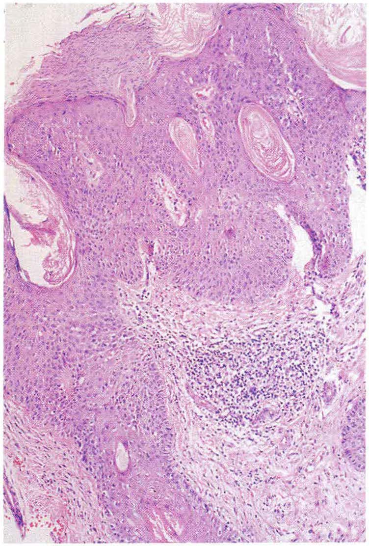
El objetivo del presente artículo es estudiar la histopatología del CEC para posteriormente analizar los factores, tanto clínicos como histológicos, que influyen en su pronóstico. Con las herramientas anteriores se hará una revisión de las diferentes modalidades terapéuticas para el manejo de CEC. Este enfoque terapéutico se basará en los factores de riesgo estudiados.
Biografía del autor/a
Ana Francisca Ramírez, Universidad del Valle
Dermatóloga, Universidad del Valle, Especialista en entrenamiento en Dermatología Oncológica, Instituto Nacional de Cancerología (INC), Bogotá D. C
Roberto Jaramillo, Universidad del Valle
Patólogo, Universidad del Valle, Cali Instituto Municipal de Investigación Médica, Barcelona, España
Luis Fernando Palma, Universidad Nacional
Dermatopatólogo, Profesor Universidad Nacional de Colombia, Bogotá D. C
Álvaro Acosta, Universidad Nacional
Dermatólogo Oncólogo, INC, Profesor Asistente Universidad Nacional, Bogotá D. C
Referencias bibliográficas
2. Acosta A. Carcinoma Escamocelular. En: Ramírez G, Patiño JF, Castro CJ. Guía Práctica Clínica en Enfermedades Neoplásicas. Bogotá, Ruecolor Ltda, 2001 :33-56.
3. Ackerman AB, Parsons L. Respect at last for solar keratosis (editorial). Dermatopathol Pract Concept 1997; 3:101-103.
https://doi.org/10.1016/S0167-2738(97)00330-5
4. Janes RE Jr. Questions to the editorial board and other authorities. What is the boundary that separates a thick solar keratosis and a thin squamous cell carcinoma?Am J Dermatopathol 1984; 6:301-306.
5. Horn TO, Moresi JM. Histology. En: Miller SJ, Maloney ME. Cutaneous Oncology, Blackwell Science 1998:481-493.
6. Hurt MA, Santacruz DJ. Tumor of the skin. En: Fletcher CDM. Diagnostic Histopathology of Tumors, Churchill Livingstone 2000: 1357-1472.
7. Kao GF. Precancerous lesions and carcinoma in situ. En: Farmer ER, Hood AF. Pathology of the Skin. Norwalk, Appleton & Lange 1990:550.
8. Ackerman B, Mones JM. Solar Keratosis? En: Ackerman B, Mones JM. Ackerman's resolving Quandaries in Dermatology, Pathology, & Dermatopathology. New York, Ardor Scribiendi 2001 :341-350.
9. Kirkham N. Tumors and cysts of the epidermis. En: Elder David. Lever's Histopathology of the Skin. Philadelphia-New York, Lippincott - Raven 1997:685-7 46.
10. Hodak E, Jones RE, Ackerman AB. Solitary keratoa cantoma is a squamous cell carcinoma: Three exampl es with metastases. Am J Dermatopathol 1993; 15:332-342.
https://doi.org/10.1097/00000372-199308000-00007
11. Cain CT, Niemann TH, Argenyi ZB. Keratoacanthoma vs squamous cell carcinoma. An immunohistochemical reappraisal of p53 protein and proliferating cell nuclear antigen expression in keratoacanthoma-like tumors. Am J Dermatopathol 1995; 17:324-331.
https://doi.org/10.1097/00000372-199508000-00003
12. Goldman GD, Leffell DJ. Cutaneous squamous cell carcinoma. En: Maloney ME, Torres A, Hoffmann T J, et al. Surgical Dermatopathology, Blackwell Science 1999: 183-223.
13. Salache S, Cheney M, Varaures M. Recognition and management of the high- risk cutaneous squamous cell carcinoma. Curr Probl Dermatol 1993; 5:141-192.
https://doi.org/10.1016/1040-0486(93)90006-H
14. Cottel WI. Perineural invasion by squamous cell carcinoma. J Dermatol Surg Oncol 1982; 8:589-600.
https://doi.org/10.1111/j.1524-4725.1982.tb00317.x
15. Goepfert H, Dichtel WJ, Medina JE. Perineural invasion in squamous cell carcinoma of the heat and neck. Am J Surg 1984; 148:542-547.
https://doi.org/10.1016/0002-9610(84)90385-4
16. Swanson PE, Fitzpatrick MM, Ritter JH, et al. lmmunohistologic differential diagnosis of basal cell carcinoma, squamous cell carcinoma and trichoepithelioma in small cutaneous biopsy specimens. J Cutan Pathol 1998; 25:153-159.
https://doi.org/10.1111/j.1600-0560.1998.tb01708.x
17. Rowe DE, Carroll RJ, Day CL. Prognostic factor for local recurrence, metastasis, and survival rates in squamous cell carcinoma of the skin, ear, and lip. J Am Acad Dermatol 1992; 26:976-990.
https://doi.org/10.1016/0190-9622(92)70144-5
18. Haas AF. Features associated with metastasis. En: Miller SJ, Maloney ME. Cutaneous Oncology. Blackwell Science 1998:500-505.
19. Baldursson B, Sigurgeisson B, Lindelbf B. Venous leg ulcers and squamous cell carcinoma: a large-scale epidemiological study. Br J Dermatol 1995; 133:571-57 4.
https://doi.org/10.1111/j.1365-2133.1995.tb02707.x
20. Steffen C. Marjolin's ulcer. Am J Dermatopathol 1984; 6:187-193.
https://doi.org/10.1097/00000372-198404000-00015
21. Dupree MT, Boyer JO, Cobb MW. Marjolin's ulcer arising in a bum scar. Cutis 1998; 62:49-51.
22. Euvrard S, Kanatakis J, Pouteil-Noble C. Comparative epidemiologic study of premalignant and malignan! epithelial cutaneous lesions developing after kidney and heart transplantation. J Am Acad Dermatol 1995; 33:222-229.
https://doi.org/10.1016/0190-9622(95)90239-2
23. Salache SJ. Features associated with recurrence. En: Miller SJ, Maloney ME. Cutaneous Oncology. Blackwell Science 1998:494-499.
24. Perez GL, Randle HW. Natural history of squamous cell carcinoma of the skin: cases report. Cutis 1995; 55:34-36.
25. Breuninger H, Schaumberg-Lever G, Holzschuk J, et al. Desmoplastic squamous cell carcinoma of the skin and vermilion surface. A highly malignan! subtype of skin cancer. Cancer 1997; 79:915-919.
https://doi.org/10.1002/(SICI)1097-0142(19970301)79:5<915::AID-CNCR7>3.0.CO;2-A
26. Liégeois NJ, Olbricht S. Squamous cell carcinoma. En: Williams H. Evidence- Based Dermatology. London, BMJ Books 2003:316-323.
27. Freeman RG, Knox JM, Heaton CL. The treatment of skin cancer: A statistical study of 1341 skin tumors comparing results obtained with irradiation, surgery, and curettage followed by electrodessication. Cancer 1964; 17:535-538.
https://doi.org/10.1002/1097-0142(196404)17:4<535::AID-CNCR2820170415>3.0.CO;2-P
28. Broadland DG, Zitelli JA. Surgical margins forexcision of primary cutaneous squamous cell carcinoma. J Am Acad Dermatol 1992; 27:241-48.
https://doi.org/10.1016/0190-9622(92)70178-I
29. Grekin RC, Salmon PJ. Surgica) Management of Local Disease. En: Miller SJ, Maloney ME, Cutaneous Oncology, Blackwell Science 1998:506-517.
30. Lawrence CM, Dahl MG, Dickinson AJ, et al. Mohs' micrographic surgery for cutaneous squamous cell carcinoma: practica! considerations. Br J Dermatol 2001; 144:186-187.
https://doi.org/10.1046/j.1365-2133.2001.03972.x
31. Werlinger KD, Upton G, Moore AY. Recurrence rates of primary nonmelanoma skin cancers treated by surgical excision corripared to electrodessication-curettage in a prívate dermatological practice. Dermatol Surg 2002; 28:1138-1142; discussion 1142.
https://doi.org/10.1097/00042728-200212000-00009
32. Kuflik EG. Cryosurgery. En: Miller SJ, Maloney ME. Cutaneous Oncology. Blackwell Science 1998:518-525.
33. Triantafyllos JF, Abrams RA. Radiation therapy. En: Miller SJ, Malloney ME. Cutaneous Oncology. Blackwell Science 1998:526-533.
34. Morton CA, Brown SB, Collins S, et al. Guidelines for topical photodynamic therapy: report of a workshop of The British Photodermatology Group. Br J Dermatol 2002; 146:552-567.
https://doi.org/10.1046/j.1365-2133.2002.04719.x
35. Greenway H. Biologic Response Modifiers. En: Miller SJ, Maloney ME. Cutaneous Oncology: Blackwell Science 1998:542-546.
36. Berg D, Otley C. Skin cancer in organ transplant recipients: epidemiology, pathogenesis, and management. J Am Acad Dermatol 2002; 47:1-17.
https://doi.org/10.1067/mjd.2002.125579
37. Mackenzie-Wood A, Kossard S, Launey J de, et al. lmiquimod 5% cream in the treatment of Bowen's disease. J Am Acad Dermatol 2001; 44:462-470.
https://doi.org/10.1067/mjd.2001.111335
38. Walsh P. Topical Chemotherapy. En: Miller SJ, Maloney ME. Cutaneous Oncology. Blackwell Science 1998:547-552.
39. Motley R, Kersey P, Lawrence C. Multiprofessional guidelines for the management of the patient with primary cutaneous squamous cell carcinoma. Br J Dermatol 2002; 146: 18-25.
https://doi.org/10.1046/j.0007-0963.2001.04615.x
40. Reschly MJ, Messina JL, Zaulyanov LL, et al. Utility of sentinel lymphadenectomy in the management of patients with high-risk cutaneous squamous cell carcinoma. Dermatol Surg 2003; 29:135-140.
https://doi.org/10.1046/j.1524-4725.2003.29035.x
Cómo citar
Descargas
Descargas
Publicado
Cómo citar
Número
Sección

| Estadísticas de artículo | |
|---|---|
| Vistas de resúmenes | |
| Vistas de PDF | |
| Descargas de PDF | |
| Vistas de HTML | |
| Otras vistas | |


